
Welcome back to the ongoing Cochlear Explorers series at Hearing International. As a quick reminder, many cells and structures in the field of anatomy are named after the researchers who initially described them, and these are referred to as Eponyms.
So far we’ve look at prominent Cochlear Explorers Alfonso Corti, Friedrich Claudius, Dieters Cells, Victor Hensen and Friedrich Claudius.
In this edition, we’ll delve into the work of Jakob Ernst Arthur Böttcher (1831 – 1889), a German pathologist and anatomist.
Böttcher’s Cells
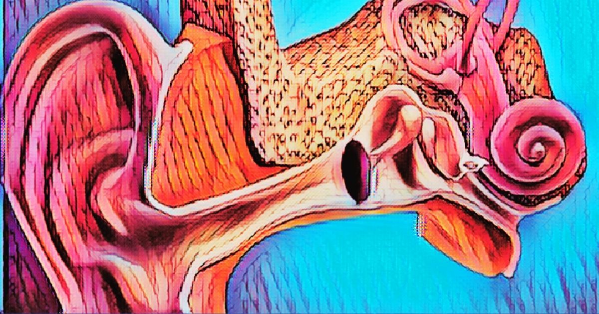
Böttcher’s cells are located on the basilar membrane, positioned below Claudius’s cells, as illustrated in the drawing on the left and the electron micrograph on the right. These cells are categorized as supportive cells for the organ of Corti and are found exclusively in the lower part of the cochlea, which is responsible for perceiving high-frequency sounds. Böttcher’s cells interlock with one another and extend microvilli into the spaces between them. Their unique structural features imply that they may play a crucial role in cochlear function.
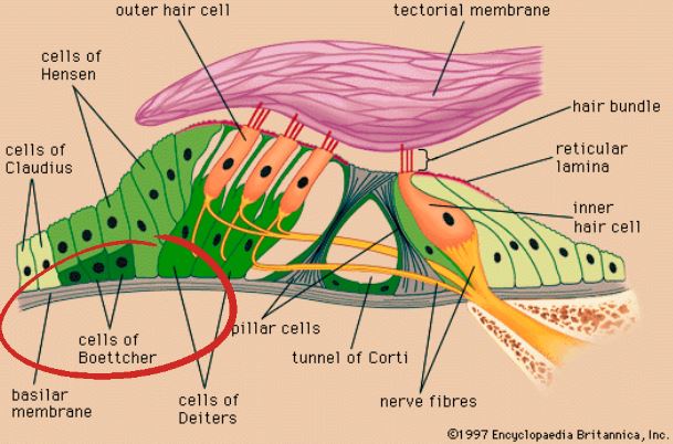
Organ of Corti. Image credit: Britannica Inc
Nitric oxide synthase (NOS) has previously been identified in various parts of the cochlea. In the cochlea, nitric oxide is believed to play a significant role in processes like neurotransmission, regulation of blood flow, and the induction of cytotoxicity under pathological conditions. However, the presence of NOS in Böttcher’s cells has been rarely reported.
Kanazawa et al. (2004) conducted a study that demonstrated the localization of NOS on Böttcher cells in rats using immunohistochemistry with a polyclonal antibody to NOS. Through observation with a light microscope using DAB staining, positive immunostaining for NOS was detected in the Böttcher cells. In immunoelectron micrographs, NOS was found in abundance in the cytoplasm of the interdigitations of these cells. This suggests that the interdigitations may play important roles in utilizing NOS, and the nitric oxide produced by Böttcher cells could influence neighboring Böttcher cells. The ultrastructure of Böttcher cells indicates that they may be active cells, serving both secretory and absorptive functions.
Arthur Böttcher
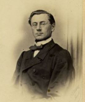
Arthur Böttcher. Image credit: Wikipedia
Arthur Boettcher, a Baltic German pathologist and anatomist, was a Russian citizen who worked in the Russian Empire during the 19th century. He was originally from Bauska, which is now in Latvia. His name is spelled as Boettcher or Böttcher in audiological literature, and this variation is a common German spelling difference. Despite the historical differences in spelling, Schacht and Hawkins suggest that it’s not a significant concern today.
Boettcher’s academic journey began in 1851 at the University of Dorpat (currently the University of Tartu in Estonia), where he obtained his doctorate in 1856. His dissertation focused on the nerve supply to the cochlea in the inner ear. Afterward, he continued his studies with visits to Germany, France, and Austria.
In 1862, he became a full professor of general pathology and pathological anatomy at Dorpat. He also served as the editor of the Dorpater Medicinische Zeitschrift (Dorpater Medical Journal) from 1871 to 1877, which was later renamed the St. Petersburg Medicinische Zeitschrift (St. Petersburg Medical Journal).
In one of his articles from 1859, Boettcher addressed various aspects of 19th-century cochlear anatomy, including the debate over whether Corti’s cells (hair cells) were connected to nerve fibers or were independent structures similar to Pacinian corpuscles. He also challenged his colleague Ernst Reissner’s description of the “Schneckenkanal” (screw channel) and membrane, asserting that he could not find them in his observations.
Boettcher’s Legacy
Boettcher is largely known for his anatomical investigations of the inner ear, particularly studies involving the structure of the reticular lamina and nerve fibers of the organ of Corti. Today his name is associated with the eponym “Boettcher cell” of the basilar membrane within the cochlea. Other anatomical terms that contain his name are:
- Boettcher’s canal, which is known today as the ductus utriculosaccularis or as the utriculo-saccular duct connecting the utricle with the endolymphatic duct.
- Boettcher’s ganglion: Ganglion on the cochlear nerve in the internal auditory meatus.
- Boettcher’s space: Also known as the endolymphatic sac; the blind pouch at the end of the endolymphatic duct.
- Charcot-Boettcher filaments: Spindle-shaped crystalloids found in human Sertoli cells. They measure 10 to 25 µm in length. Named in conjunction with neurologist Jean-Martin Charcot (1825-1893).
Boettcher received the prestigious Karl von Baer Prize of the St Petersburg Academy of Sciences (the forerunner of the Russian Academy of Sciences) for his work on the structure of the labyrinth within the ear. He quit working in approximately 1877 at about age 45 as he became ill. After 12 years of illness, he died at the age of 57 in August 1889.
Next week we look into another Cochlear Explorer, Friedrich-Christian Rosenthal
About the author
Robert M. Traynor, Ed.D., is a hearing industry consultant, trainer, professor, conference speaker, practice manager and author. He has decades of experience teaching courses and training clinicians within the field of audiology with specific emphasis in hearing and tinnitus rehabilitation. He serves as Adjunct Faculty in Audiology at the University of Florida, University of Northern Colorado, University of Colorado and The University of Arkansas for Medical Sciences.
**this piece has been updated for clarity. It originally published on July 7, 2014


