
In the November 2015 PCAST guidance recommendations, it was noted that “in the near future, people could check their hearing using automated hearing tests available online” and could use computer & smartphone interfaces that “allow adaptive self-fitting by devices in response to user needs. Custom earbuds and configurations could be made routinely by 3D printing.”
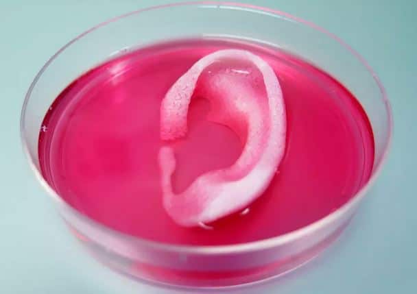
Additive manufacturing, better known as 3-D printing is a process used to make a three-dimensional object, using successive layers of material that form under computer control to create an object. In addition to using 3D printing for custom earmolds and earbuds, researchers at the Wake Forest Baptist Medical Center have shown that living tissue structures can be 3-D printed, including ear, bone and muscle, which may be used to replace tissue on human patients.
No Longer Science Fiction

Dr. Anthony Atala
In a story first reported by medical writer, Stacy Lawrence, Dr. Anthony Atala, director of the Wake Forest Institute for Regenerative Medicine and senior author on the study in a statement was quoted, “This novel tissue and organ printer is an important advance in our quest to make replacement tissue for patients. It can fabricate stable, human-scale tissue of any shape. With further development, this technology could potentially be used to print living tissue and organ structures for surgical implantation.”
The research team published a paper in the journal Nature Biotechnology showing that they were able to 3-D print human-scale tissues and then effectively implant them in mice to result in vascularized, functional tissue. Next, the scientist attempted to replicate this 3-D process using human tissue. Their early results in this area demonstrate that these 3-D printed tissues have the correct size, strength and function for use in humans.
According to Lawrence, it took researchers 10 years to develop the Integrated Tissue and Organ Printing (ITOP) System that printed these tissues at the Institute for Regenerative Medicine.
Unlike traditional 3-D printing that operates via jetting, extrusion or laser-induced forward transfer, the ITOP system deposits both biodegradable, plastic-like material to for tissue shape and water-based gels that contain the particular cells.
The researchers made tissue structures with vessel-like structures that were smaller than 200 microns (0.007 inches) to help the cells to survive. In the study, they created a baby-sized ear structure of 1.5 inches that was implanted on a mouse and showed signs of vascularization one and two months after implantation as well as maintained its shape.
https://youtu.be/jvzu9MXC7e0


