
Descriptions of the ear canal, and understanding not just its anatomy, but also its termination (tympanic membrane/eardrum), have significant implications for the taking of ear impressions, and also for any device that couples an auditory device to the ear. There are certain issues that have always bothered me relative to this topic, so I decided to post this blog to see what kind of dander I could raise, especially in Part II of this series. First, some basics.
Ear Canal Length
The ear canal is terminated by the tympanic membrane (TM, or ear drum) – Figure 1. Measurements of the ear canal length vary substantially – with adult averages from approximately 19 to 34 mm from the aperture (opening) of the ear canal to the TM.
Textbooks generally specify the average length at 25 mm, but references and measurement procedures for providing this length are vague. Most descriptions seem to assume this length and do not document the reference. Unfortunately, some of the references used consist of an n of one. While the length range appears to be rather wide, there are some accepted explanations. In that which follows, all comments relate to adult human ears.
Terminal Measurement Point
First, it is not clear what part of the TM constitutes the terminal measurement location. The TM is not vertical (perpendicular to the ear canal), but instead angles rather dramatically, something not ordinarily shown in renderings of the ear anatomy (Figure 1). Figure 2 illustrates a more realistic rendering of the TM angle in the ear canal, and Figure 3 provides additional information about the ear canal angle when viewed from above. Data from Salvinelli, et al. (Figure 7) indicates that the angle is about 43 degrees to the central canal axis, and about 19 degrees anterior-inferior, with the back upper part being closest to the opening of the ear canal {{1}}[[1]] Salvinelli, F., Maurizi, M., Calamita, S., D’Alatri, L., Capelli, A., and Carbone, A. 1991. Scandinavian Audiology, 20, 253-256[[1]]. Similar angles are reported by Smelt, et al., 1988) of 47 degrees for the central canal axis, and 17 degrees for the anterior-inferior {{2}}[[2]] Smelt, G., Hawke, M. and Proops, D. 1988. Anatomy of the external ear canal: a new technique for making impressions. The Journal of Otolaryngology, 17, 249-253[[2]].
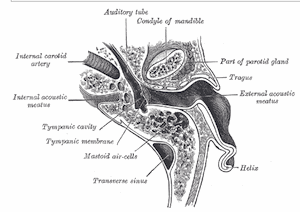
Figure 3. Right ear tympanic membrane as viewed from above, showing that the posterior portion is closest to the aperture of the ear canal.
Figure 1. The ear canal terminating at the Tympanic membrane. Artist’s rendering showing the tympanic membrane fairly vertical.
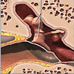
Figure 2. Frontal view of the tympanic membrane showing a more realistic angle of its position in terminating the ear canal.
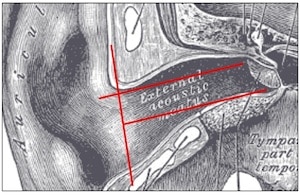
Figure 4. Ear canal length, showing that the superior portion of the TM is closest to the external environment. Both red lines from the aperture to the TM are the same length.
Ear canal length is reported in Figures 5 and 6. Staab {{3}}[[3]] Staab, W. Deep canal Hearing aids: Theory and practice,” ASHA Miniseminar, New Orleans, Louisiana, November 1994[[3]] and Oliviera {{4}}[[4]] Oliviera, R. The dynamic ear canal, Chapter 6, The Human Ear Canal, Ballachanda (Ed), Singular Publishing Group, Inc., San Diego, 1995 pp. 84-111[[4]] (Figure 6) specify the TM measurement point as the furthest distance on the TM from the aperture. Other studies are assumed to use the center of the TM as the terminal measurement, but this is not clear. As illustrated in Figure 4, where the measurement is terminated on the TM can lead to a 3-5 mm difference in ear canal length (red lines into auditory canal are both the same length).
Cadaver Vs. Living Ear Measurements
A second consideration relates to measurements of ear impressions made from cadaver versus live ears. It has been explained to expect some shrinkage with cadaver ears, and, if true, the length will be assumed shorter as a result. The ear canal lengths of cadaver ears in Figure 5 show less of a range than those from live ears in Figure 6. From the measurements shown, it appears that cadaver ears have a length of approximately 22.5 to a little over 25 mm (although where the measurement was made to on the TM was not specified).
For ear canal measurements from live persons, the length appears to be from about 19 to 34 mm in length, without taking gender differences into consideration (Figure 6).
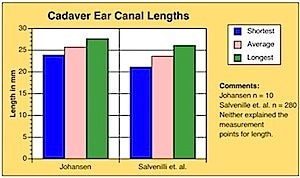
Figure 5. Ear canal lengths based on ear impressions from 208 cadaver ears. Redrawn from Salvinelli, et. al., 1991, and Johansen, 1975.
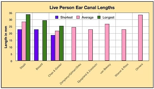
Figure 6. Ear canal lengths reported for live persons.
Comments related to Figure 6:
- Staab measurements were taken from ear impressions. n = 38. Measurement to longest part of TM
- Bezold {{6}}[[6]]Bezold, F. Die Corrosions-Anatomie des Ohres, 1882[[6]]. Measurements were taken from ear impressions. n = 20. Measurement termination position not identified
- Chan & Giesler used an optical method {{7}}[[7]] Chan, J., and Giesler, C. 1980. Estimation of eardrum acoustic pressure and of ear canal length from remote points in the canal. Journal of the Acoustical Society of America, 87(3), 1237-1247[[7]]. The n was not identified. Measurement termination to top of TM
- Zemplenyi, Gilman, and Dirks used an optical method {{8}}[[8]] Zemplenyi, J., Gilman, S., and Dirks, D. 1985. Optical method for measurement of ear canal length. Journal of the Acoustical Society of America, 78, 2146-2148[[8]]. The n was not identified. The umbo was the measurement target.
- Djupesland and Zwislocki {{9}}[[9]] Djupesland, G., and Zwislocki, J. 1972. Sound pressure distribution in the outer ear. Scandinavian Audiology, 4, 197-203[[9]] used an acoustic method. n = 7.
- Von Békésy {{10}}[[10]] von Békésy, G. 1960. Experiments in Hearing, New York: McGraw-Hill[[10]] used an acoustic method. The n was not identified, but much of his work consisted on an n of 1.
- Wiener and Ross {{11}}[[11]] Wiener, G., and Ross, D. The pressure distribution in the auditory canal in a progressive sound field, J. Acoust. Soc. Amer., 1946, 18, 401-408[[11]] used an acoustic method. n = 6 (12 ears?).
- Oliveira used an MRI image. n = 1.
Ear Canal Length Gender Differences
A third consideration is that female ear canals tend to be shorter than those of males by about 1 to 3.7 mm (Salvinelli et. al., 1991; Chan and Giesler, 1980; Zemplenyi, Gilman, and Dirks, 1985). The largest population size reporting on this difference was that of the cadaver measurements by Salvinelli, et al. who showed female ear canals to be 2.7 mm shorter on average than for males. It would appear to logically apply that difference to ear impressions taken from living adults. Staab and Bezold, who reported on the largest number of measurements from living persons, did not break out gender difference in the measurements.
Measurements of ear canal dimensions were not simple to make in the past. As a result, until recently, information was not available that described the configuration or the dimensions well. What follows is a partial list of measurement procedures that have been used. Procedures are posted that make volume and other measurements as well, even though this presentation is interested primarily in the length of the ear canal with its termination at the TM.
Best Estimates of Ear Canal Length
The best estimates for the overall adult population (male and female) seems to be as identified below. The next blog will separate the male from the female population based on the available information.
- 27 mm when measured to the bottom of the TM
- 25 mm when measured to the umbo (center of the TM)
- 23 mm when measured to the upper part of the TM
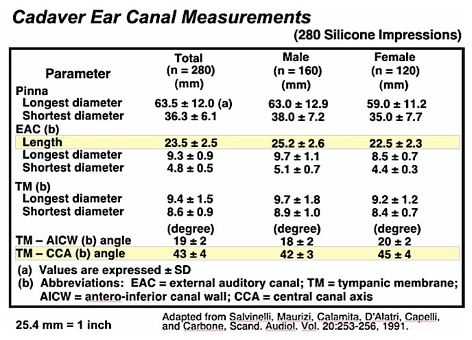
Figure 7. Cadaver ear canal measurement details from Salvinelli, et. al., 1991.
Ear Canal Measurement Methods
A variety of approaches have been used to measure the ear canal for length, diameter, volume, etc. Even though this presentation is interested primarily in length measurements, it is helpful to be exposed to other measurement procedures used to gain additional ear canal information.
1. Physical Measurements.
a. Direct physical measurements of ear canal impressions. This has the advantage of providing information about the configuration as well as the dimensions. The difficulty with this method is that making impressions of the complete ear canal, including the imprint of the tympanic membrane, is no simple task. As a result, most measurements made this way are made from ear impressions of cadaver ears. The primary question relates to the use of cadaver ear impressions as accurate measurements of living ear canals. A large number of complete ear impressions have been made on live individuals for the Philips XP Peritympanic hearing aid in the mid-1990s. Except for a few remaining in my possession, essentially all others have been destroyed.
b. Another physical measurement (of volume, but not of configuration) has involved the use of complete ear impressions and use of a calibrated syringe filled with a 60% alcohol solution. The volume is a function of the distance the ear impression is inserted into the syringe solution.
2. Optical Measurements.
This is a noninvasive procedure that has been used to measure the length of the ear canal, using a microscope. The microscope is focused on the tympanic membrane. It is then refocused on an earplug in the ear that terminates at the aperture of the ear canal. The focus measurement difference provides information about the length.
3. Radiological Measurements.
These noninvasive procedures involve CT and MRI scans. The CT is best for identification of bony structure, whereas the MRI is used best for measurement of soft tissues. A primary objection to the use of these techniques is the exposure to radiation.
4. Acoustic Measurement Method (mathematical description)
This procedure measures the sound pressure at 2+ locations in the ear canal. A broadband signal is used to make the SPL measurements. The second step involves the determination of the area using a linear system of equations and executing a simple iterative algorithm. Or, another way is to use 1/4 wave theory and look at the maxima and minima to calculate the length.
5. Immittance Measurements:
Commercially-available instruments are available that measure the canal volume from the tip of the earpiece placed in the ear to the tympanic membrane. Unfortunately, they do not provide measurements of the entire ear canal, and, they overestimate the actual volume.
6. 3-Dimensional scanning is a recent approach that is being used to measure ear canal dimensions to provide for the fabrication of earmolds, replacing ear impression taking procedures. To the best of my knowledge, these have not yet been used to measure the ear canal length through the TM.
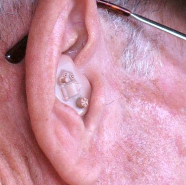
*featured image courtesy flckr


Nice approach to something most of us take for granted. I look forward to the discussion of the TM, especially it’s strength against ear mold impression material.
Mike: I will try to keep it interesting.
I would like to think that the 3D scanning of the ear canal will be able to give the clinician a more accurate measurement of the ear canal length.
If this is not something that is incorporated in the device at present then I think it should be considered.