
HANNOVER, GERMANY — In our inner ear, there are two different types of sensory cells that are responsible for hearing. A German research team has now identified the molecular switch for the formation of these inner and outer hair cells and thus found an important building block for the treatment of hearing loss.
The inner and outer hair cells develop before birth from a common type of precursor cells. Which factors control the different development was unknown for a long time. A research team led by Professor Dr. Andreas Kispert and Dr. Mark-Oliver Trowe from the Institute of Molecular Biology at the Hannover Medical School (MHH) has now found the key and demonstrated how it controls the process: The gene Tbx2 acts like a switch to determine whether inner or outer hair cells are formed. The work was supported by the German Research Foundation (DFG) and has been published in the renowned journal Nature Communications.
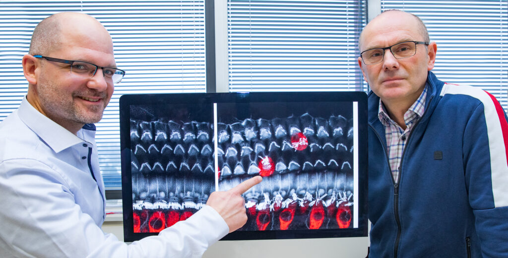
Dr. Mark-Oliver Trowe (left) and Professor Dr. Andreas Kispert with the image of inner (red) and outer (gray) hair cells from the inner ear of the mouse; Copyright: Karin Kaiser / MHH.
Vibration transducers and sound amplifiers
More than 400 million people worldwide are affected by hearing loss. The most common is sensorineural hearing loss, in which the hair cells responsible for hearing are destroyed.
From birth, we have about 15,000 hair cells in each ear. They are located in the inner ear in the cochlea. 3500 of them belong to the inner hair cells. They are the actual sensory cells and convert the vibrations triggered by external sound into electrical signals in the inner ear. These reach the brain via nerve pathways, where the auditory impression is created. The outer hair cells act as a mechanical amplifier and improve the sensitivity of hearing.
Once destroyed, the hair cells can no longer regenerate and the hearing ability decreases. “This happens, for example, due to ageing processes, excessive noise or even the intake of certain medications,” says Dr Trowe, head of the study. Scientists around the world are working to develop therapies that can help renew the auditory sensory cells. “But for this, it is imperative to understand which genes control the formation of hair cells during embryonic development.”
Tbx2 regulates development into inner and outer hair cells
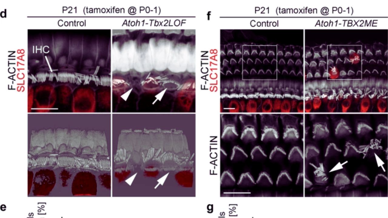
In their investigations, the research team focused on the gene Tbx2. It contains the information for a so-called transcription factor. Like an on-off switch, this protein regulates whether certain genetic information on the DNA should be read or not. “We studied the cochlea in the mouse model and found that Tbx2 is active exclusively in the inner hair cells,” says Professor Kispert. If Tbx2 was specifically switched off in the precursor cells, no more inner hair cells formed. If, on the other hand, it was switched on in all precursor cells, only inner hair cells were formed. The importance of the molecular switch for the allocation of hair cells was also shown at a later stage of development.
“If Tbx2 is deactivated in the inner hair cells, they transform into outer hair cells. Conversely, activation of Tbx2 in the outer hair cells causes them to convert to inner hair cells.”
–Dr. Andreas Kispert
Since the inner ear of mice and humans is similar, the researchers assume that the regulatory pathways are also transferable. The identification of Tbx2 as a molecular switch for the formation of inner hair cells is therefore an important building block for the development of regenerative therapies for the treatment of hearing loss.
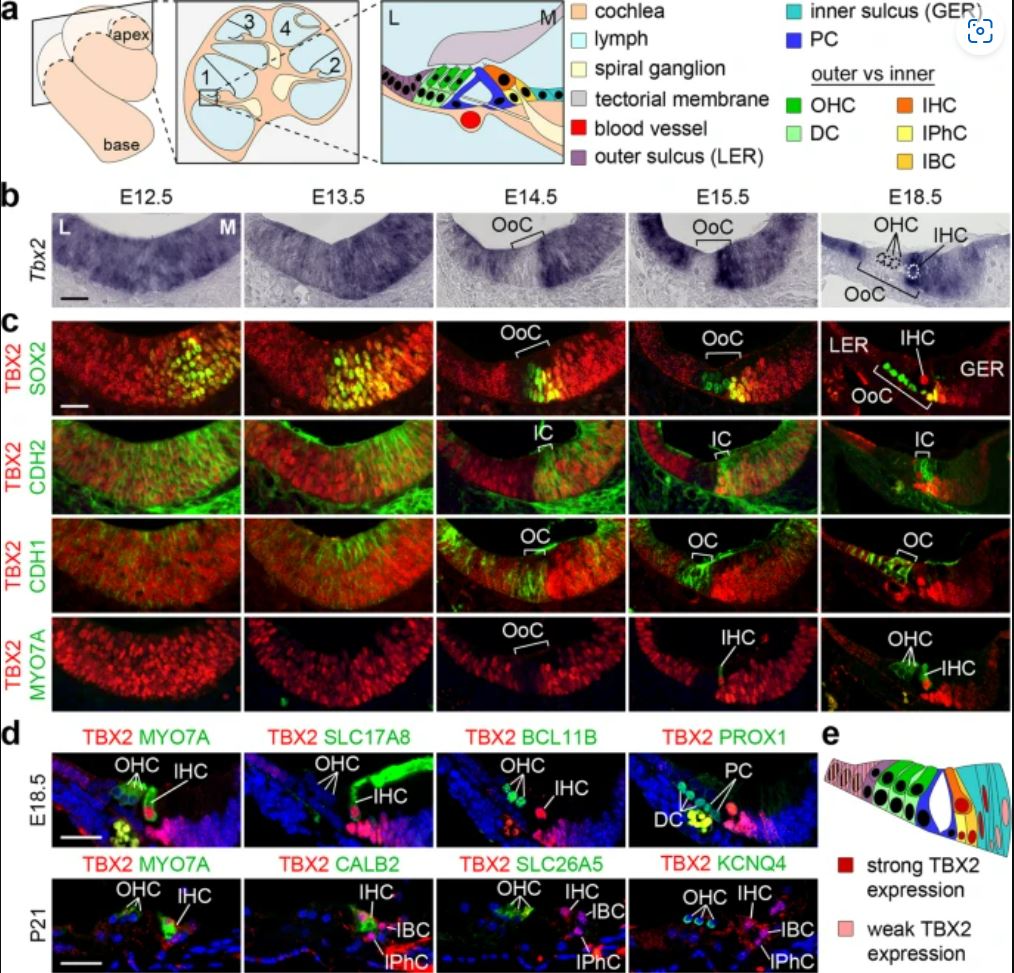
a Scheme showing the basal (1) and the apical (4) turn of the mature cochlea, a cross section through the cochlea, and a magnification of the basal turn with the cellular organization in and around the organ of Corti (OoC): with Deiters’ cells (DCs) and OHCs in the outer compartment (OC, shades of green), inner phalangeal cells (IPhCs), inner border cells (IBCs) and IHCs in the inner compartment (IC, shades of yellow) separated by pillar cells (PCs) that form the tunnel of Corti. Laterally, the OoC is flanked by cells of the outer sulcus, and medially by cells of the inner sulcus. GER greater epithelial ridge (primordium of the inner sulcus), L lateral, LER lesser epithelial ridge (primordium of the outer sulcus), M medial. b–d Expression of Tbx2 mRNA and TBX2 protein on sections of the developing cochlea of wildtype embryos. b RNA in situ hybridization analysis of Tbx2 expression in the prosensory epithelium of the cochlea at E12.5 and E13.5 as well as in the developing OoC between E14.5 and E18.5. Dotted circles mark the nucleus of the three OHCs and of one IHC in the OoC at E18.5. c, d Co-immunofluorescence analysis of expression of TBX2 with markers for differentiation (SOX2: pro-sensory cells/supporting cells; MYO7A: hair cells) and compartmentalization (CDH2: inner; CDH1: outer) from E12.5 to E18.5 (c) and with markers for all hair cells (MYO7A), IHCs (SLC17A8, CALB2), OHCs (BCL11B, SLC26A5, KCNQ4), DCs and PCs (PROX1) at E18.5 and P21 (d). n = 3 for each assay at each stage. Scale bars: 30 µm. e Scheme of TBX2 expression (red nuclei) in the OoC at E18.5. The color code is the same as in a.
- The original paper “TBX2 specifies and maintains inner hair and supporting cell fate in the Organ of Corti” with first author Dr. Marina Kaiser can be found here.
Source: MHH, Nature Comm


