Middle Ear Muscles and Meniere’s
Last week, I mentioned that a study advocated severing the tendons to the tensor tympani and stapedius muscles as a treatment for Meniere’s disease. I admitted that I was not familiar with this treatment and decided to look into it. Here is what I found:
For this to make any sense, we must do a quick review of the function of these muscles. A rudimentary description of the hearing process involves sound waves impacting the tympanic membrane (the eardrum), which in turn transmits vibration to the 3 ossicles. The ossicles are the 3 bones connecting the tympanic membrane to the inner ear. These are known as the malleus, incus, and stapes. The stapes bone is connected to the oval window, which is a membranous connection to the inner ear. The stapes works almost like a piston, pumping back and forth against the oval window, creating waves in the fluid housed within the inner ear. The primary function of this system is to amplify sounds that arrive at the tympanic membrane and transition them to fluid waves within the cochlea, which eventually become signals transmitted to the brain.
The tensor tympani and stapedius muscles provide a protective role, in that they are stimulated and contract in response to loud, potentially damaging noise. The tensor tympani muscle is attached to the malleus, while the stapedius muscle is attached to the stapes. When the brain detects a sound that is loud enough to potentially damage the inner ear, these muscles contract. The tensor tympani muscle reduces vibration at the eardrum, and the stapedius muscles dampens the vibration of the stapes. This quickly, but not immediately, reduces the loudness of the sound.
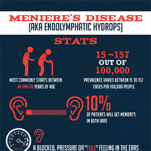
Theoretically, these muscles, even when they are not contracted, may have some influence over the freedom of movement of the ossicles. As it relates to Meniere’s disease, the theory has been proposed that Meniere’s disease can occur as a result of endolympahatic hydrops ( AKA cochlear hydrops), or an excess of inner ear fluid. It has been proposed that by disconnecting these muscles from the ossicles, the oval window can better accommodate increased fluid pressure within the inner ear.
According to Loader et al, “…middle ear muscles would increase the inner ear pressure in cochlear hydrops of Meniere’s disease, by pressing the ossicular chain in the opposite direction to the bulging of the endolymphatic hydrops.
This seems plausible, considering that the rise in inner ear pressure has three main routes of escape: the vestibular aqueduct, the round window, and the oval window (upon which the stapes is mounted). In analogous fashion, if the stabilizing function of the middle ear muscles is interrupted by tenotomy, the ossicle chain can be moved laterally more freely, which results in a bulging of the tympanic membrane laterally. The ossicular chain is no longer actively pressed against the oval window and therefore the pressure on inner ear structures is not heightened even further.”
This theory was tested (sort of) by a group out of Austria who published a report in 2011 demonstrating a reduction in Meniere’s symptoms after undergoing the tenotomy procedure, which is described as “The stapedius and tensor tympani tendons were cut with a neurectomy knife. No damage to the bony ossicular chain was caused.” The authors acknowledge that Meniere’s symptoms generally improve over time regardless of treatment, but they report that unlike other methods (including the natural course of the disease) improvements in vertigo attacks were noted immediately. They also acknowledge that there was no control group in this study, stating, “we opted not to include a control group because this study did not aim to prove one surgical technique superior to another, but solely to show the immediate and long term effects of tenotomy.”
This treatment is not part of the typical treatment ladder for Meniere’s, but until we have a better understanding of the mechanics of Meniere’s, and a reliable treatment, no theory should be abandoned without consideration.


Having dealt with quite a few ear surgeons over the years, one of the less common observations they shared with me was that simply opening the middle ear sometimes seemed to result in a lessening of Meniere’s symptoms. Maybe just the trauma? Maybe something else? Maybe just anecdotal. Certainly interesting, especially in light of this posting.
It could be a placebo. Trials have been been done where they cut people and sewed them up but didn’t do anything. Yet people claimed their surgery worked.
An interesting study. But why lose a critical inhibitory function to reduce vertigo, when we know that Meniere’s symptoms reduce with time ? Losing inhibition and control of incoming sounds will almost certainly exacerbate existing hearing loss, but making it noise induced.