Approximately one year ago, I embarked on an exploration of the most efficient and cost-effective methods for assessing patients experiencing acute dizziness. This investigation took the form of a comprehensive five-part blog series titled “Acute Vertigo: Could It Be A Stroke?” Within this series, I underscored a crucial point: the prevalent imaging techniques often employed in the Emergency Room setting tend to be significantly less efficacious than relatively simple screening protocols.
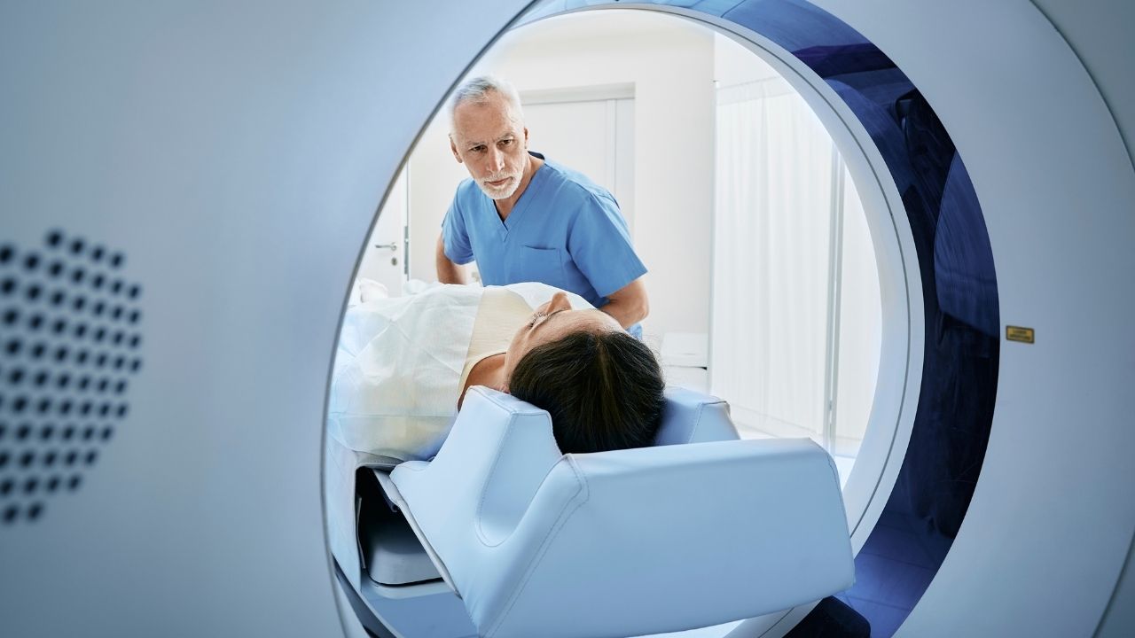
Recently, a study surfaced in the ED Management Journal, revealing the outcomes of a three-year research endeavor conducted at Henry Ford Hospital in Detroit. This study, entitled “Study: reconsider the use of CT scans for patients who present with dizziness,” examined the diagnostic efficacy of CT (Computerized Tomographic) scans administered to patients with complaints of dizziness, as requested by emergency department physicians. The findings were striking, with less than one percent of these CT scans yielding any substantial diagnostic information. Astonishingly, the financial cost associated with these scans, performed over a span of three years within a single emergency room, amounted to nearly one million dollars.
It is worth noting that in the few instances where positive findings were observed on the CT scans, the dizziness was consistently accompanied by symptoms like headaches or other neurological deficits. This correlation highlights the potential utility of CT scans for a particular subset of cases.
Value of CT Scans for Dizziness: Limited
However, when it comes to the more prevalent complaints of dizziness, such as lightheadedness, positional vertigo, or isolated dizziness, the value of performing a CT scan appears to be very limited. These types of dizziness are unlikely to yield significant insights through CT imaging. As such, the study illuminates the need for a nuanced and targeted approach to diagnostic imaging, taking into account the clinical context and the specific nature of the patient’s symptoms.
This study’s revelations prompt a broader discussion on the balance between diagnostic precision and resource optimization in healthcare. The allure of advanced imaging techniques often overshadows the potential benefits of streamlined screening protocols.
Equally significant is the sensitivity of CT scans in identifying cerebellar stroke, a concern paramount to the ordering physician. A noteworthy consideration arises due to the common occurrence of normal CT scans within the initial hours after an acute ischemic stroke, rendering a standard CT scan ineffective in excluding the possibility of a cerebellar stroke. This limitation poses a potential challenge to accurate diagnosis, as up to half of cerebellar stroke cases could remain undetected if reliant solely on CT scans for assessment.
As many as 50% of cerebellar stroke patients may be missed if diagnosis is dependent on CT scanning.
Bottom line, CT scans ordered for dizziness hardly ever provide pertinent diagnostic information, and may miss a high percentage of the very few with potentially worrisome intra-cranial pathologies.
References:
- Study: reconsider the use of CT scans for patients who present with dizziness. ED Management Journal, 2012, April:24(4):45-47
- Simmons et al. Cerebellar infarction: comparison of computed tomography and magnetic resonance imaging. Ann Neurol. 19, 291-293
About the author
 Alan Desmond, AuD, is the director of the Balance Disorders Program at Wake Forest Baptist Health Center, and holds an adjunct assistant professor faculty position at the Wake Forest School of Medicine. He has written several books and book chapters on balance disorders and vestibular function. He is the co-author of the Clinical Practice Guideline for Benign Paroxysmal Positional Vertigo (BPPV). In 2015, he was the recipient of the President’s Award from the American Academy of Audiology.
**this piece has been updated for clarity. It originally published on June 4, 2012


