It was fitting that this series began last week with Alfonso Corti, since most of the Cochlear Explorers in this series are named for components of his famous Organ of Corti.
The origins of anatomical terminology can be traced back to the early days of humanity when our ancestors first started naming different parts of the human body. The use of these terms has proliferated over time, to the extent that the field of anatomy is now at risk of becoming overwhelmed by its own nomenclature.
It’s astounding that there are more than 50,000 names assigned to approximately 5,000 anatomical structures.
This problem, which was recognized in 1917, has likely only intensified over the years. Many cells and structures are named after the researchers who first described them, a practice referred to as “eponyms.” While one might assume that having structures named after them was a career highlight in the 19th century, the early anatomists who described cochlear components didn’t use this as a means to achieve fame or immortality. Instead, these eponyms arose incidentally and evolved slowly.
 In the 19th century, it was common to identify anatomical structures by the discoverer’s first name, such as referring to “the cells that Claudius described.” This approach was convenient because the original authors often had limited understanding of the cells’ roles and precise structures, making it challenging to assign precise anatomical names. Naming cells this way likely contributed to confusion regarding cochlear anatomy.
Who Was Friedrich Claudius?
This edition of Cochlear Explorer spotlights Friedrich Matthias Claudius (June 1, 1822 – January 10, 1869). In a 1855 paper titled “Bemerkungen uber den Bau der hautigen Spiralleiste der Schnecke” (Remarks on the structure of the membranous spiral strip of the snail) published in a German Zoology journal, Friedrich Claudius detailed what would later be known as the “Cells of Claudius.”
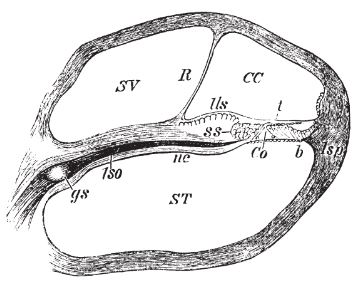
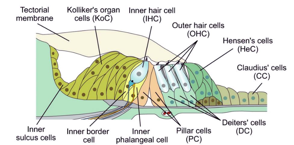
The upper diagram of the cochlea depicts the Cells of Claudius on the far right side, while in the lower diagram showcasing various eponymous cochlear structures, they are located on the left. These cuboidal supporting cells are part of the Organ of Corti within the Cochlea, extending from Hensen’s cells to the spiral prominence epithelium, forming the Outer Sulcus. They come into direct contact with the endolymph of the scala media and are sealed through tight junctions that prevent endolymph from flowing between them. Boettcher cells are situated directly below the cells of Claudius in the lower cochlear turn.

Diagram of the cochlear epithelium in young postnatal mice showing inner and outer hair cells and various types of supporting cells. Click here for more details. Credit: Vélez-Ortega et al./Nature Communications
Limited information is available about Friedrich Matthias Claudius. He was born in Lubeck, Germany, in 1822, around the same time as Corti’s birth in Italy. He grew up in the historic part of Lubeck, close to the Trave River, which was connected to the Elbe River by the Elbe Lubeck Canal. Lubeck is located on the Baltic coast, not far from Hamburg.
While Friedrich was related to the famous 18th-century German poet Matthias Claudius (1740-1815), no surviving images of him exist. What we do know is that Friedrich earned his doctorate in Zoology from the University of Göttingen in 1844. In 1849, he was appointed to the Zoological Museum of Kiel University and later became a professor and director of the anatomical institute at the University of Marburg. Beyond these details, not much else is known about the individual who first described the Cells of Claudius.
Join us next week for a closer look at another Cochlear Explorer—Hensen’s Cells.
About the author

Robert M. Traynor, Ed.D., is a hearing industry consultant, trainer, professor, conference speaker, practice manager and author. He has decades of experience teaching courses and training clinicians within the field of audiology with specific emphasis in hearing and tinnitus rehabilitation. He serves as Adjunct Faculty in Audiology at the University of Florida, University of Northern Colorado, University of Colorado and The University of Arkansas for Medical Sciences.
**this piece has been updated for clarity. It originally published on June 17, 2014


Please continue this. I am sure it will be of benefit to thousands. Medicine on a whole is packed with Eponyms. One hears of the ganglion cells of Gasser, ligament of Poupart, foramen of Vesalius, ligament of Sir Astley Paston Cooper, Copula of His, Criminal Nerve of Grassi. Maybe you will come across something on Grassi. I have actually searched for information on Grassi but have come up empty handed. Blessings on you!