
KANSAS CITY, MO — While humans can regularly replace certain cells, such as those in the blood and gut, most other tissues in the body do not naturally regenerate. For example, when the tiny sensory hair cells in the inner ear are damaged, the result is often permanent hearing loss, deafness, or balance disorders. In contrast, animals like fish, frogs, and chicks can regenerate these cells with ease.
Now, scientists at the Stowers Institute for Medical Research have identified how two distinct genes guide the regeneration of sensory hair cells in zebrafish. This discovery deepens our understanding of regenerative biology in zebrafish and may help guide future studies focused on hearing loss and regenerative medicine in mammals, including humans.
“Mammals such as ourselves cannot regenerate hair cells in the inner ear,” said Stowers Investigator Tatjana Piotrowski, Ph.D., the study’s co-author. “As we age or are subjected to prolonged noise exposure, we lose our hearing and balance.”
Gene Roles in Cell Division and Regeneration
New research from the Piotrowski Lab, published in Nature Communications on July 14, 2025, investigates how cell division is regulated to promote hair cell regeneration while maintaining a stable pool of stem cells. Led by former Stowers Researcher Mark Lush, Ph.D., the team found that two different genes involved in cell division regulate two key types of sensory support cells in zebrafish. These findings may help researchers explore whether similar processes could be triggered in human cells.
“During normal tissue maintenance and regeneration, cells need to proliferate to replace the cells that are dying or being shed — however, this only works if there are existing cells that can divide to replace them. To understand how proliferation is regulated, we need to understand how stem cells and their offspring know when to divide and at what point to differentiate.”
–Tatjana Piotrowski, Ph.D.

Graphic illustration of zebrafish neuromast showing where the progenitor and stem cells reside. Image credit: SIMC
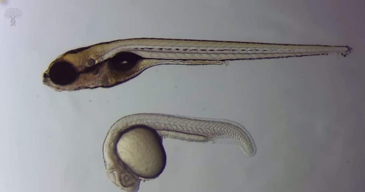
Zebrafish provide a valuable model for regeneration research. Sensory organs called neuromasts, arranged in a line from head to tailfin, help zebrafish detect water motion. Each neuromast resembles a garlic bulb, with hair cells sprouting from the top and surrounded by support cells. These structures closely resemble the hair cells in the human inner ear.
Because zebrafish are transparent during development and possess accessible sensory organs, researchers can directly visualize and genetically manipulate individual neuromast cells. This allows detailed investigation into the mechanisms of stem cell renewal, progenitor cell proliferation, and hair cell regeneration.
“We can manipulate genes and test which ones are important for regeneration,” said Piotrowski. “By understanding how these cells regenerate in zebrafish, we hope to identify why similar regeneration does not occur in mammals and whether it might be possible to encourage this process in the future.”
Distinct Genes, Distinct Functions
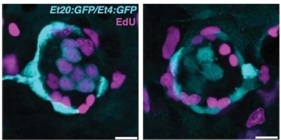
When the proliferation gene responsible for progenitor populations is disrupted (right), progenitors regenerate hair cells through direct differentiation in the absence of proliferation. Wild type neuromast is on the left. Image credit: SIMC
Two main populations of support cells contribute to hair cell regeneration within the neuromast: active stem cells located at the edges and progenitor cells near the center. These cells divide symmetrically, enabling ongoing production of new hair cells while preserving the stem cell pool. Using sequencing techniques, the researchers identified two distinct cyclinD genes, each active in only one of the two populations.
Genetic alterations revealed that each cyclinD gene independently regulates the cell division of its respective population. When the gene responsible for progenitor cell proliferation was disrupted, those cells ceased dividing but still formed hair cells through direct differentiation.
“When we rendered one of these genes non-functional, only one population stopped dividing,” said Piotrowski. “This finding shows that different groups of cells within an organ can be controlled separately, which may help scientists understand cell growth in other tissues, such as the intestine or blood.”
Remarkably, when the gene typically used by stem cells was engineered to function in progenitor cells, their ability to divide was restored — demonstrating the genes’ cell-type specificity.
David Raible, Ph.D., a professor at the University of Washington who studies the zebrafish lateral line sensory system, commented on the significance of the findings. “This work illuminates an elegant mechanism for maintaining neuromast stem cells while promoting hair cell regeneration. It may help us investigate whether similar processes exist or could be activated in mammals.”
Because cyclinD genes also regulate cell division in human tissues such as the gut and blood, the team’s work may have broader implications beyond hearing.
“Insights from zebrafish hair cell regeneration could eventually inform research on other organs and tissues, both those that naturally regenerate and those that do not,” said Piotrowski.

Additional authors of the study include Ya-Yin Tsai, Shiyuan Chen, Daniela Münch, Julia Peloggia, Ph.D., and Jeremy Sandler, Ph.D.
This research was funded by the National Institute on Deafness and Other Communication Disorders of the National Institutes of Health (NIH) (award: 1R01DC015488-01A1), the Hearing Health Foundation, and institutional support from the Stowers Institute for Medical Research. The content is solely the responsibility of the authors and does not necessarily represent the official views of the NIH.
Reference:
Lush, M.E., Tsai, YY., Chen, S. et al. Stem and progenitor cell proliferation are independently regulated by cell type-specific cyclinD genes. Nat Commun 16, 5913 (2025). https://doi.org/10.1038/s41467-025-60251-0
About the Stowers Institute for Medical Research
Founded in 1994 through the generosity of Jim Stowers, founder of American Century Investments, and his wife, Virginia, the Stowers Institute for Medical Research is a non-profit, biomedical research organization with a focus on foundational research. Its mission is to expand our understanding of the secrets of life and improve life’s quality through innovative approaches to the causes, treatment, and prevention of diseases.
The Institute consists of 20 independent research programs. Of the approximately 500 members, over 370 are scientific staff that include principal investigators, technology center directors, postdoctoral scientists, graduate students, and technical support staff. Learn more about the Institute at www.stowers.org and about its graduate program at www.stowers.org/gradschool.
Source: SIMC, Nature Communications