
A new study, published this week in JAMA Otolaryngology-Head & Neck Surgery, has demonstrated the complementary diagnostic capabilities of middle ear optical coherence tomography (OCT) and computed tomography (CT) in diagnosing various middle ear conditions.
The study, which examined three different patient cases, suggests that while OCT and CT each have their strengths and limitations, their fusion can provide unparalleled insight into both soft tissue and bony structures of the middle ear.
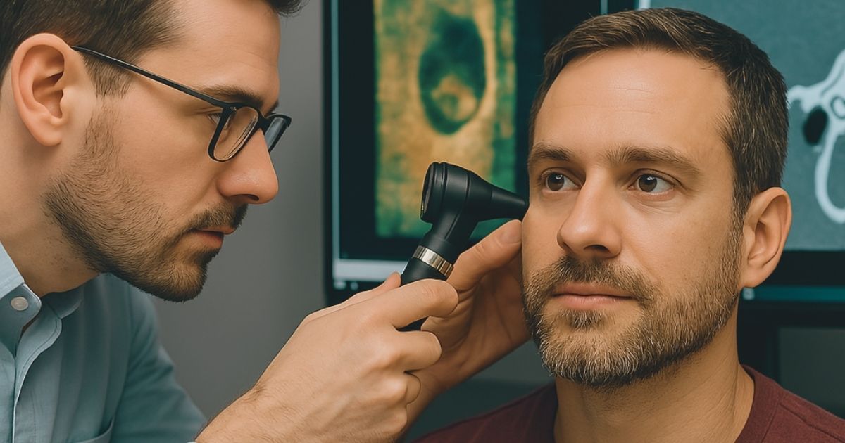
Complementary Roles of OCT and CT in Middle Ear Diagnostics
The study included three patients with distinct middle ear conditions, providing OCT and CT images for comparison. By using OCT’s high-resolution imaging for soft tissue and CT’s ability to visualize bony structures, the researchers were able to generate co-registered images, revealing the potential for enhanced diagnostics in otology. The researchers emphasized that while OCT cannot replace CT due to its limited field of view (FOV) and inability to penetrate thick bone, it provides a detailed look at soft tissue features such as adhesions and cholesteatoma that are difficult to detect in CT scans.
This ability to capture high-resolution soft tissue images in real time without ionizing radiation makes OCT a promising tool in middle ear diagnostics, particularly in settings like pediatric care or early screenings.
The case series included a normal middle ear, a traumatic middle ear injury, and a cholesteatoma-affected ear. The OCT images provided new insights into the soft tissue conditions that were not as clearly visible in CT scans. For example, in the traumatic ear injury case, OCT highlighted the displacement of the ossicles, which CT images failed to detect due to the lack of visibility of the tympanic membrane. Similarly, in the cholesteatoma case, OCT effectively differentiated the cholesteatoma from surrounding tissues, showing the potential for OCT to assist in identifying pathology that may be missed in traditional imaging.
The Promise of Real-Time Imaging and Future Applications

Researchers suggest that combining OCT and CT images could provide a more complete picture of the middle ear and improve diagnostic accuracy
The study’s findings indicate that OCT, with its ability to visualize soft tissue in real time and without the use of ionizing radiation, can complement traditional CT scans in clinical otology. The researchers suggest that combining OCT and CT images could provide a more complete picture of the middle ear and improve diagnostic accuracy, especially in complex cases such as trauma or chronic ear infections. Though OCT currently faces limitations such as a smaller FOV and sensitivity to obstructions, its real-time imaging capability and detailed soft tissue visualization hold great potential for future use in middle ear diagnostics and surgery.
As this technology evolves, OCT may become an essential tool in combination with traditional imaging technologies, offering a non-invasive, high-resolution alternative to better diagnose and monitor middle ear diseases. The study calls for further development to address the challenges posed by OCT’s limitations and looks forward to the potential integration of OCT with other technologies, such as OCT Doppler vibrometry, which can provide additional functional information.
In conclusion, the fusion of OCT and CT imaging represents an exciting advancement in otology. With further refinement, OCT could become an integral part of the diagnostic toolkit for middle ear disorders, offering real-time imaging capabilities that enhance both the accuracy and efficiency of patient care.
Citation:
- Wang JCouvreur FFarrell JD, et al. Fusion of Middle Ear Optical Coherence Tomography and Computed Tomography. JAMA Otolaryngol Head Neck Surg. Published online April 03, 2025. doi:10.1001/jamaoto.2025.0043
**Readers interested to learn more about OCT and its usefulness in the ENT/Audiology clinic can check out this recent interview with Dr. John Oghalai: