
For the first time, researchers have demonstrated that terahertz (THz) imaging can be used to visualize internal structures of the cochlea with micron-level resolution—without damaging the delicate tissue. The study, conducted by a team led by Kazunori Serita at Waseda University in Japan and published in Optica, could pave the way for new diagnostic tools to detect hearing loss and ear-related conditions earlier and more accurately.
“Hearing relies on the cochlea, a spiral-shaped organ in the inner ear that converts sound waves into neural signals,” said Serita.
“Although conventional imaging methods often struggle to visualize this organ’s fine details, our 3D terahertz near-field imaging technique allows us to see small structures inside the cochlea without any damage.”
Terahertz Imaging: A New Approach
Terahertz radiation, which lies between microwaves and infrared light on the electromagnetic spectrum, is well-suited for biological imaging. It is non-ionizing, meaning it poses no harm to tissues, and it can pass through bone while offering high sensitivity to variations in hydration and tissue composition. This makes it uniquely suited to studying the cochlea, which is encased in bone and filled with lymphatic fluid.
However, traditional THz imaging tools have limitations. Lenses used to focus THz waves typically generate beams several millimeters wide—too large to resolve the cochlea’s microscopic structures. To overcome this, the researchers developed a near-field terahertz point source just 20 microns in diameter by using a nonlinear optical crystal. This allowed them to achieve much higher spatial resolution than previously possible.
“Until now, there was no way to observe the internal structure of the cochlea non-destructively with high resolution,” said Serita. “A key innovation in our work was the use of a nonlinear optical crystal to generate terahertz waves from 1560-nm near-infrared light. This was crucial for our imaging technique.”
From 2D to 3D: Visualizing the Cochlea’s Interior
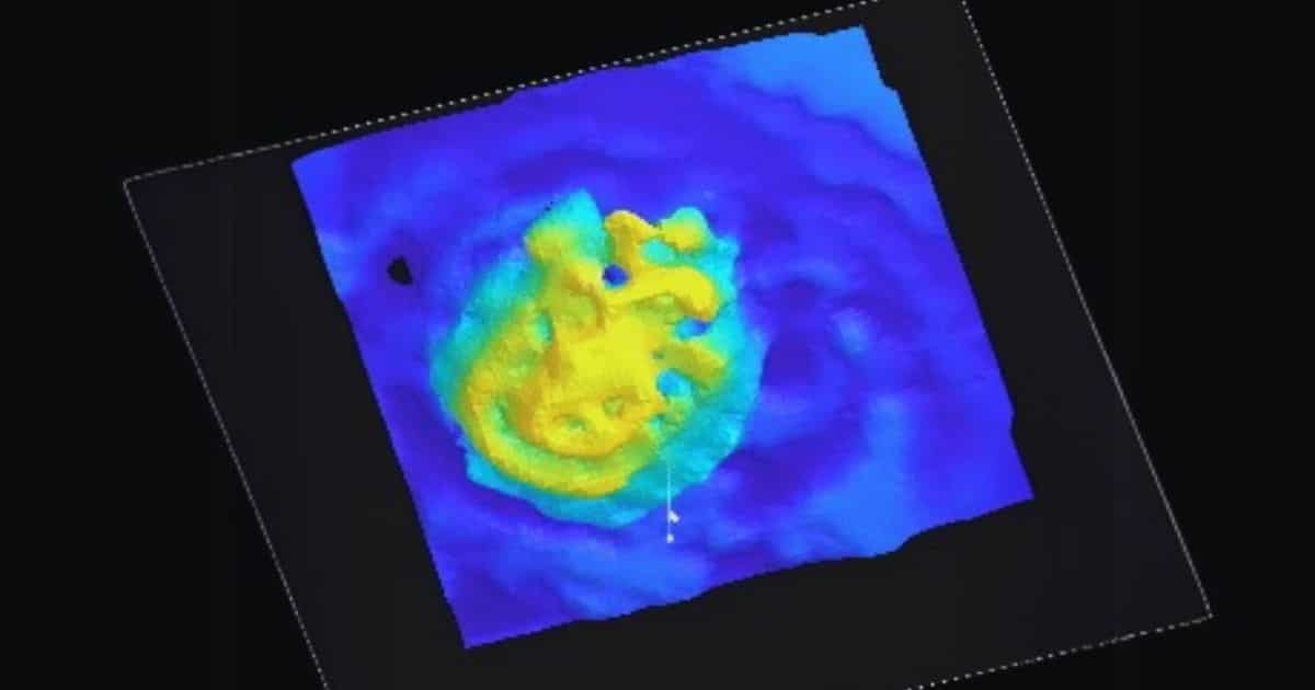
To validate the method, the team conducted imaging tests on two extracted and dried mouse cochleae—one left empty and another filled with metal. Clear visual differences confirmed that the terahertz radiation was reaching and interacting with internal structures. Using a time-domain imaging setup, they then extracted internal details and applied an unsupervised learning algorithm to highlight features within the cochlea.
The imaging setup also enabled 3D time-of-flight imaging and reconstruction, successfully visualizing part of the cochlear duct—the spiral canal essential to the cochlea’s function. These 3D reconstructions showed that the system could accurately map inner structures in high detail.
“With further development, this technique could lead to a new diagnostic method for ear diseases that have been difficult to diagnose until now. It has the potential to enable on-site diagnosis of conditions like sensorineural hearing loss and other ear disorders. It might also be useful for early detection of hearing impairments, allowing earlier treatment and better outcomes.”
Toward Clinical Applications
The current imaging tests were conducted on dried tissue samples, but the research team is now working to adapt the technology for use in live biological environments. Because the cochlea is deeply embedded and filled with fluid, future iterations of the system will need to be miniaturized and powered by stronger THz sources to enable sufficient penetration and imaging depth.
Serita was inspired to explore this approach after conversations with Takeshi Fujita, a co-author from the Department of Otolaryngology–Head and Neck Surgery at Kobe University, who described the challenges of imaging the cochlea in clinical settings. “That got me thinking — maybe terahertz imaging could help solve these issues,” said Serita. “We decided to collaborate and explore this idea together. The big question was whether we could visualize the tiny internal structures of the cochlea without causing any damage.”
The researchers say the technology could eventually be integrated into otoscopes or endoscopes, enabling non-invasive in vivo imaging not only for cochlear diagnostics but also potentially for early cancer detection and other medical applications.
Citation:
- L. Zheng, H. Chen, T. Fujita, A. Kakigi, N. Allen, H. Murakami, M. Tonouchi, K. Serita, “Three-dimensional terahertz near-field imaging evaluation of cochlea,” 12, 437–445 (2025). DOI: 10.1364/OPTICA.543436.
About Optica Publishing Group
Optica Publishing Group is a division of the society, Optica, Advancing Optics and Photonics Worldwide. It publishes the largest collection of peer-reviewed and most-cited content in optics and photonics, including 18 prestigious journals, the society’s flagship member magazine, and papers and videos from more than 835 conferences. With over 400,000 journal articles, conference papers and videos to search, discover and access, our publications portfolio represents the full range of research in the field from around the globe.
About Optica
Optica is an open-access journal dedicated to the rapid dissemination of high-impact peer-reviewed research across the entire spectrum of optics and photonics. Published monthly by Optica Publishing Group, the Journal provides a forum for pioneering research to be swiftly accessed by the international community, whether that research is theoretical or experimental, fundamental or applied. Optica maintains a distinguished editorial board of more than 60 associate editors from around the world and is overseen by Editor-in-Chief Prem Kumar, Northwestern University, USA. For more information, visit Optica.
Source: Optica