For people with severe to profound hearing loss, cochlear implants can provide access to sound that may significantly improve communication and quality of life. However, outcomes vary widely among recipients, and understanding which patients are most likely to benefit remains an important clinical question.
A research project led by Yingying Wang at the University of Nebraska–Lincoln examined how brain activity and sensory integration may influence speech perception outcomes in cochlear implant (CI) users. The three-year study explored how neuroimaging techniques could help predict which candidates may experience the greatest benefit from implantation.
Cochlear implants are complex electronic devices designed for individuals with severe to profound hearing loss. Unlike hearing aids, which amplify sound, cochlear implants bypass damaged portions of the inner ear and directly stimulate the auditory nerve to provide a representation of sound signals. Their effectiveness can depend in part on how well the auditory nerve and associated brain networks function.
Examining the Brain’s Role in Cochlear Implant Outcomes
The study was conducted through collaboration between researchers at the University of Nebraska–Lincoln, the University of Nebraska Medical Center (UNMC), and Ohio State University.
Wang serves as resident faculty in the Center for Brain, Biology and Behavior and directs the Neuroimaging for Language, Literacy and Learning Lab. She is also affiliated with the Nebraska Center for Research on Children, Youth, Families and Schools.
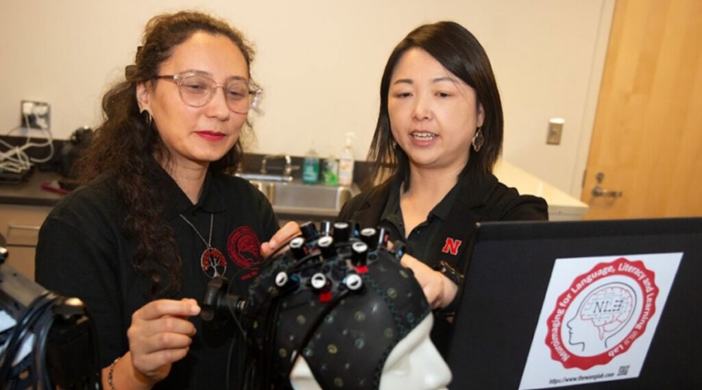
Yingying Wang, associate professor of special education and communication disorders, discusses how researchers evaluate potential candidates for cochlear implantation. Image credit: University of Nebraska–Lincoln
The research examined how factors such as age and hearing thresholds influence the connectivity and network efficiency of the brain. The team also investigated the role of a measure known as the visual analog of temporal envelope, which reflects the brain’s ability to integrate sensory information.
According to the researchers, this measure appeared to support speech perception for cochlear implant users in moderately noisy environments.
These findings highlight the potential value of neuroimaging tools—including resting-state functional magnetic resonance imaging (fMRI) and functional near-infrared spectroscopy—in studying how the brain processes sound following implantation.
Tracking Brain Changes Before and After Implantation
The project examined brain activity in cochlear implant candidates both before and after surgery. Prior to implantation, participants underwent neuroimaging to identify brain regions responsive to speech sounds and to confirm whether the auditory nerve remained intact.
Following surgery, the research team monitored changes in brain activity and speech perception during follow-up visits. The goal was to better understand why some cochlear implant recipients achieve stronger speech perception outcomes than others.
Wang compared the adaptation process to adjusting to a new pair of eyeglasses, noting that the brain must learn to interpret new sensory input.
“For people who have been deaf for years, this region has often been recruited for vision or touch. Now sound signals are rushing in again, and the brain needs to relearn how to process them.”
The project was funded by the National Institute on Deafness and Other Communication Disorders. In addition to Wang, the research team included co-principal investigator Michelle Hughes, professor of special education and communication disorders; Jonathan Hatch, an otolaryngologist and assistant professor at UNMC who performed cochlear implant procedures; and Hongying (Daisey) Dai, an associate professor at UNMC who served as the project’s statistician.
Recruitment proved challenging during the study. Twelve adults with severe-to-profound hearing loss completed pre-surgical evaluations, but only five participants completed post-surgical assessments.
Because of the limited sample size, Wang is seeking additional federal funding to expand the research. The proposed follow-up study would include additional cochlear implant candidates recruited in Columbus, Ohio, while the Nebraska research site would focus on comparison participants.
Potential for Future Interventions
Looking ahead, Wang hopes that further research could help identify pre-surgical interventions that improve cochlear implant outcomes by supporting the brain’s ability to adapt to auditory input.
“By examining the brain’s neuroplasticity, we may be able to determine pre-surgery interventions to benefit patients,” Wang said.
Understanding how the brain adapts to cochlear implant stimulation may ultimately help clinicians better predict patient outcomes and develop strategies to improve speech perception after implantation.
References
Ellsworth, C. T., et al. (2025). Age-Related Hearing Decline and Resting-State Networks. American Journal of Audiology. DOI: 10.1044/2025_aja-25-00025.
Gao, Z., et al. (2025). Visual Analog of Temporal Envelope Benefits Speech Processing in Cochlear Implant Users: A Pilot Functional Near-Infrared Spectroscopy Study on the Associations Between Audiovisual Benefit, Listening Environment, and Peripheral Neural Health. Ear & Hearing. DOI: 10.1097/aud.0000000000001709.
Source: UNL, AJA, E&H