
Welcome back to the ongoing Cochlear Explorers series here at Hearing International. Just a quick reminder, many cells and structures are named after the researchers who first described them, and these are known as Eponyms.
This week, our focus is on Otto Friedrich Karl Deiters (1834-1863), a German neuroanatomist.
Deiters Cells
First, let’s understand what and where Deiters cells are located. Deiters cells are a type of supportive cell. Along with Hensen’s cells and Claudius cells, they provide support to the outer hair cells within the cochlear structure. Deiters cells span from the basilar membrane to the reticular lamina, and when combined with pillar cells and outer hair cells, they play a crucial role in defining the micro-architecture of the organ of Corti.
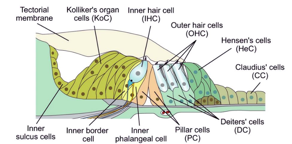
DIAGRAM OF THE COCHLEAR EPITHELIUM IN YOUNG POSTNATAL MICE SHOWING INNER AND OUTER HAIR CELLS AND VARIOUS TYPES OF SUPPORTING CELLS. CLICK HERE FOR MORE DETAILS. CREDIT: VÉLEZ-ORTEGA ET AL./NATURE COMMUNICATIONS
A recent study by Parsa, Webster, and Kalinec in 2012 made an interesting discovery. They found that the complete detachment of Deiters cells reveals an elliptical imprint on the top surface of the basilar membrane. This imprint consists of a smaller central structure with a very smooth surface surrounded by a rougher area. This suggests the presence of two different anchoring junctions.
These previously unidentified morphological features of Deiters cells may have a significant impact on the mechanical response of the organ of Corti.
Otto Friedrich Karl Deiters
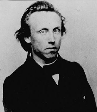
Otto Friedrich Karl Deiters. Image credit: Wikipedia
Otto Deiters, born in Bonn, Germany in 1834, is known within the scientific community for his significant contributions, particularly in the field of neuroanatomy. One of his most notable discoveries is the “nucleus of Deiters,” also referred to as the “lateral vestibular nucleus,” and “Deiters’ cells,” which are associated with the outer hair cells in the inner ear’s cochlea.
Tragically, Deiters’ promising career was cut short when he passed away at the age of 29 in 1863 due to typhoid fever. Much of his work was posthumously edited and published by his colleague and professor, Max Schultze, a microanatomist from the University of Bonn, where Deiters had spent most of his brilliant but brief career.

University of Bonn
Deiters made notable contributions to the study of the brain and spinal cord. In the 1860s, he provided the most comprehensive description of a nerve cell known at that time. He identified the cell’s axon, which he termed an “axis cylinder,” and its dendrites, which he referred to as “protoplasmic processes.” He also proposed that dendrites must merge to form a continuous network.
One of his famous publications, “Untersuchungen Über die Lamina Spiralis Membranacea” (Investigations over the Lamina Spiralis Membranous), was published in 1860. In this work, he described the supporting cells of the hair cells. Deiters’ original drawings from this book are shown on the right.
Considering the limited tools available at the time, such as the relatively rudimentary microscopes, his ability to identify and outline these structures was a remarkable feat.
Next Week’s Cochlear Explorer is German Pathologist Arthur Boettcher.
About the author

Robert M. Traynor, Ed.D., is a hearing industry consultant, trainer, professor, conference speaker, practice manager and author. He has decades of experience teaching courses and training clinicians within the field of audiology with specific emphasis in hearing and tinnitus rehabilitation. He serves as Adjunct Faculty in Audiology at the University of Florida, University of Northern Colorado, University of Colorado and The University of Arkansas for Medical Sciences.
**this piece has been updated for clarity. It originally published on July 1, 2014