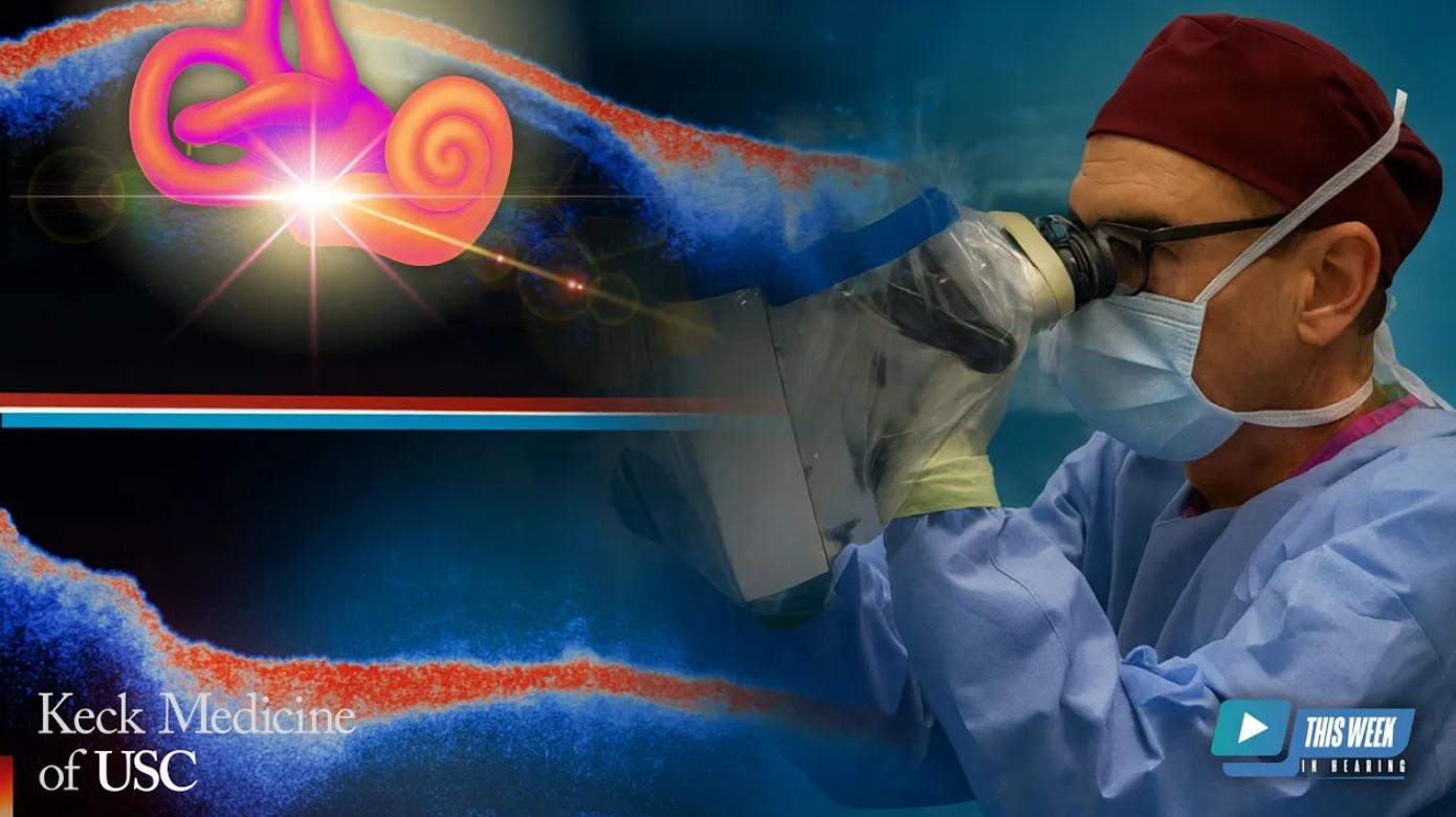
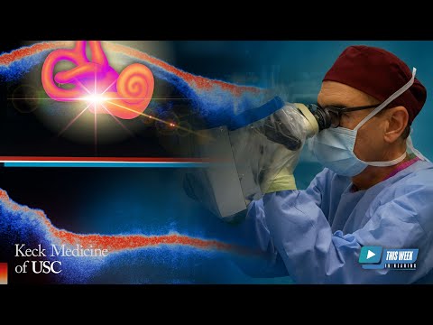
For the first time, optical coherence tomography (OCT) is being used to study the human inner ear directly in patients. In this follow-up conversation, Dr. John Oghalai, Professor and Chair of the Caruso Department of Otolaryngology at USC, shares the latest advances in this groundbreaking research. Dr. Oghalai describes how his team has transitioned from animal studies to human imaging, uncovering new insights into conditions such as Meniere’s disease and vestibular schwannoma.
By visualizing endolymphatic hydrops in real patients, this work moves closer to a future where handheld OCT devices could help audiologists and clinicians diagnose and monitor inner ear disorders in everyday practice.
The discussion explores both the promise and current challenges of developing practical, non-invasive imaging tools—technology that may one day transform care for hearing loss, tinnitus, and balance disorders.
- Learn more about the work being conducted in Dr. Oghalai’s lab here
Full Episode Transcript
Speaker 1: Welcome to This Week in Hearing. Hello, I’m Bob Traynor, your host for this episode, and today is a very special day here at This Week in Hearing, because we’re going to look at some further research from from our colleague Dr. Jon Ogulay. And he’s the professor and chair at the Caruso Department of Otolaryngology within the Keck School of Medicine at the University of Southern California. Dr. Ogulay is back to present further research with optical coherence tomography, and this time, it has to do with human research, which is quite a progression from where he had been in the past. And so thanks so much for being with us again, Jon, here at This Week in Hearing.
Speaker 2: Oh, thanks, Bob. It’s an honor to be asked back to, to visit your show once again.
Speaker 1: Well, you know it looks like the last episode that you and I did, even though we had a few complications was the most watched episode of This Week in Hearing for the last year or so. So so I’m really excited to see what you have actually done in terms of progress on your research and but before we get
Speaker 2: I just, I got to speak up just so the audience knows exactly what the, the challenges were at the last, … little
Speaker 1: Well, I deserved that, I know. So.
Speaker 2: So, for those of you who didn’t recognize it, the first time we did the whole one-hour interview, but we had forgotten to press the record button. And so we had to do a second one-hour interview where we could re-record the whole thing. But it went very well. I had a nice practice run. So I do appreciate that, Bob.
Speaker 1: Well, today, I don’t think we’ll do any practice runs. We’ll just kind of get into it a little bit here, Jon. And so but before we get going, there may be some people who didn’t really hear the, the one we did last time, and could you just give us a brief orientation to how you got to, into doing research in hearing as an otolaryngologist, as well as a little bit about the optical coherence tomography that you’re using in your research?
Speaker 2: Well, thanks. Yeah. So I’m a clinician scientist, and about half of my time is in the clinic and operating rooms and some administrative duties. And then the other half is in the research lab. And you know, I started doing this in medical school, and I’ve been doing it throughout residency and, and my career. And we initially were using rodents, a lot of guinea pigs and mice rats to some degree. But, you know, as a clinician, I really wanted to bring the work to help my patients. And so I’m excited about this work because it is a translation of some of the work that we did in animal models, and now we can start to use it to understand human disease.
Speaker 1: Wow.
Speaker 2: So, so that’s, that’s what’s exciting to me about this work.
Speaker 1: Okay. So, can you, can you give us an idea of how you transitioned with optical coherence tomography from going from the animals? Now, I understand you’re working with humans in this area.
Speaker 2: Yeah. You know, so optical coherence tomography, it’s a very common clinical tool. It’s used in ophthalmology to image the retina and also portions of the lens and even the cornea of the eye. And so most ophthalmologists will have an OCT system in their clinic. And essentially what it does is it gives you a depth profile, kind of like an ultrasound, but instead of shooting in ultrasonic waves, it shoots in light waves. And then some of the light wave is reflected back, and based on the pattern of reflected light, you can get a depth profile. And if you, if you scan the light beam across the tissue, you get a full 3D view of the tissue. And we wanted to bring that to bear to see, well, would it work in the ear? And we started with animal models because, you know, one problem with the ear is it’s deep inside the base of the skull, surrounded by otic capsule bone. That bone is the densest bone in the body, and it’s really hard to get in there because if you were to make a hole through that bone and enter the fluids of the inner ear, there’s a very good chance you’re going to damage hearing and the very delicate processes that are going on there related to it. And, and so the nice thing about, we thought, about OCT is if we could send the light in through the bone, then it could reflect back and give us a non-invasive picture inside the inner ear. And we started using mice. Mice have small cochlea and small vestibular systems, but the bone is also thinner over it. And it’s also a very good lab animal to use. We know there’s a lot of hearing loss models that have been made in mice. And in fact, for most of hearing research, that’s how we understand you know, human disease, is with a mouse model of the of the inner ear pathology. So at any rate, we did a lot of work in mice and we, you know, were using OCT. We could image in the cochlea, we could play sounds, we can measure how the basilar membrane, the tectoral membrane, the hair cells are, are vibrating in response to the sound. And we’ve done a lot of the preliminary work on that. And one of the interesting things we found, well, I guess it was now it’s been about eight or nine years ago-… is that when mice are exposed to a loud sound, like a a noise exposure for a couple of hours, or even just a sudden blast exposure, they develop endolymphatic hydrops. The, the, the endolymph, the fluid within scala media becomes greater. And we could see that with OCT very clearly as a distention of Reissner’s membrane. And that kind of happened transiently, and then after a few hours, it would kind of, you know, go down back to normal again. And we recognize that that’s probably why, after you go to a loud rock concert, you feel kind of a sense of fullness in your ears and you have some hearing loss, and it’s the endolymphatic hydrops that you’re feeling. And so, we can see that in our mice, and that’s exciting because patients with Meniere’s disease, we, you know, at least we know that… We think that they have endolymphatic hydrops too. The data from that mostly comes from postmortem temporal bone histopathology, meaning the patient dies, you take out their temporal bone and you fix it and you slice it up and look at it under the microscope. Now, there’s a lot of artifact that can happen with this kind of temporal bone histopathology. It’s a great science, but there’s a lot of artifact that can happen and so it’s always been a question of, well, is endolymphatic hydrops really the, you know part of the pathologic process of Meniere’s disease? So, that’s what this work was designed to do, was to take the… You know, we, we know in a mouse we could see endolymphatic hydrops. We think humans with Meniere’s disease have endolymphatic hydrops. You know, let’s look in the humans and see if they really do.
Speaker 1: Yeah. So, so how did the humans do?
Speaker 2: Well, they did pretty good. So we, you know, our OCT systems, you can buy commercial OCT systems. You can buy them for clinical purposes. Of course, those are most… Those are made for the eye for the most part. And you can buy research systems that are really, you know, almost, I mean, would be hard to use for anything but an experiment on an animal. And so, we kinda hand-build our own systems rather than purchasing them and so we built one that can fit on the front of an operating microscope. So, when I’m in surgery, drilling on a mastoid on a patient with ear disease, then this thing is, is right there and we can bring it in and, and look with it, and we can In this case, we decided to look into the lateral semicircular canal and the posterior semicircular canal, because they’re really right there when you’re doing a mastoidectomy. And so, we brought it in and we could look in there. So, we looked at three different groups of patients. So, the, the control group were patients that have chronic ear disease, meaning they have, you know, chronic mastoiditis, a cholesteatoma, chronic otitis media. At any rate, for whatever reasons, their, their cochlea has normal bone lines, but they have a conductive hearing loss usually. And so, we figured that’d be a control for endolymphatic hydrops, because we wouldn’t expect any other inner ear disease. Then we had a group of patients with Meniere’s disease. That was our second group. And those patients were undergoing endolymphatic shunt surgery. We don’t do that very often, but when we do it, it’s a very convenient way to look at their semicircular canals, ’cause we have to do a mastoidectomy to get to the the vestibular aqueduct and endolymphatic sac. And then the third group of patients were patients that have vestibular schwannoma, that we were doing a translabyrinthine approach to the cerebellar pontine angle. And the reason we picked that group, well, there’s two reasons. Number one, part of the surgery is drilling through the balance canals, through the, the semicircular canals to get, you know, to the tumor, and so we knew we were gonna be able to thin the bone over it. We thought, “Well, that might make it easier to see now with our system.” The other thing is that there’s actually some data out there suggesting that these patients have endolymphatic hydrops. This is based on some MRI data where they’re able to get some assessment of the endolymph volume. There’s also findings of increased protein in the inner ear, which we see by MRI. We can see it on the fluid assessment of the MRI. And so, we thought, “Well, there’s a good chance we’ll be able to see endolymphatic hydrops in those, you know, those patients too.” So, those were our three groups, the control patients with chronic otitis media, the experimental groups, one with Meniere’s disease and one with vestibular schwannoma. And we imaged through the bone with our system, and we could, we could detect the, the membranous labyrinth, which is the thin membrane separating the endolymph from the perilymph. And so, we took these images and then we analyzed, well, what is the cross-sectional area of the endolymph and what is the, the cross-sectional area of the perilymph? And so, we create a ratio, the ratio of the endolymph to perilymph, and we have a normal, a ratio in normals, and then we have ratios for those patients with Meniere’s disease, and we have a endolymph to perilymph ratio for those with vestibular schwannoma. … and it turns out that the Meniere’s disease patients definitely had endolymphatic hydrops. They had much higher endolymph to perilymph ratio. And surprisingly, the vestibular schwannoma patients were even higher. It was just amazing. Yeah.
Speaker 1: And so this built on the research from before as well as moving forward then to, to to almost a diagnostic assessment of individuals. So you could almost… Further research, I suspect, will allow ENT and possibly even audiology someday to look at these differences and be able to tell. Certainly, Meniere’s disease has always been a mystery of course, in all of our careers, I’m sure. And and to be able to do that is a definite move forward in the diagnosis of the disorder itself.
Speaker 2: Yeah. Thank you. I, I agree. I think… I mean, the downside of this technique is right now it needs to be done in the operating room.
Speaker 1: Yeah.
Speaker 2: And so, you know, there’s not a lot of indication for doing that for a diagnostic purpose. That would be that would be a pretty big undertaking.
Speaker 1: Sure.
Speaker 2: But the fact that we can see this endolymphatic hydrops in humans using OCT is a, a very strong feasibility argument for continuing to work on this technology to build a, like, a handheld otoscope version that you could use in a clinic and just look down the ear canal.
Speaker 1: Wouldn’t that be cool?
Speaker 2: Yeah.
Speaker 1: Oh, geez. for something that you couldn’t… you could say, well, it’s kind of like a Meniere’s disease syndrome, or it’s kind of a, kind of a… It’s kind of this kind of a syndrome, go from that, to hold the otoscope up and say, “Well, it looks like you have this.” You know, it’s quite a, quite an advancement.
Speaker 2: Well, we’re getting there. I mean, actually, I have it right here. I can show it to you.
Speaker 1: Oh, great.
Speaker 2: So this is what we got right now.
Speaker 1: Okay. Wow.
Speaker 2: Yeah. You just hold it up there, and you can look in people’s ear, and there’s a wire. You see there’s some yellow?
Speaker 1: Uh-huh.
Speaker 2: Those are fiber optic cables carrying the light.
Speaker 1: Okay.
Speaker 2: And then some electronic cables, and they go to a computer.
Speaker 1: Oh, okay.
Speaker 2: And so it’s kind of neat because there’s a there’s a video camera in there, so you get a really good picture of the, the ear canal and the tympanic membrane, you know, just like you would with an with a microscope or with an endoscope.
Speaker 1: You know,
Speaker 2: But then you also get an OCT image.
Speaker 1: … I was gonna say, it started with brick phones, right? And then we
Speaker 2: Yeah.
Speaker 1: on to this, this, this, and this. So
Speaker 2: Yeah.
Speaker 1: … over time, your concept can be digested into something that could be used by, by, by a, a hometown ENT guy that’s looking for these kinds of disorders
Speaker 2: Or, or frankly, I mean, our goal is to build these in a stable enough system, not with the cables, kind of obvious
Speaker 1: Oh, yeah. Sure.
Speaker 2: … that we can deploy it to our audiology clinics, because I actually see audiologists using this more than… I mean, ENT might use it, but I think audiology would use it.
Speaker 1: Well, like, like a lot of things ENTs start using things, and then we find ways that it can be put in the hands of capable audiologists to facilitate a, an assessment that they can report back to the, to the to the ENT for what’s going on here. So almost like eCochs started out that way in some of those things.
Speaker 2: Mm-hmm.
Speaker 1: … so again now the As you were doing this, you had to remove a lot of that bone ’cause it’s so dense, I I assume, to
Speaker 2: Yeah.
Speaker 1: … really see some of these things, but maybe over time, they could figure out a way to penetrate that bone without having to do anything.
Speaker 2: Well, in the semicircular canals, we don’t have to thin the bone.
Speaker 1: Okay.
Speaker 2: We can see right through the natural bone.
Speaker 1: Okay.
Speaker 2: In the cochlea, it’s a little more challenging. The bone is about the same thickness, but if you’re looking down the ear canal, there’s two other things you have to go through, not just the otic capsule bone. You have to go through the eardrum. That’s one.
Speaker 1: Yeah.
Speaker 2: That’s… It’s thin, but it does take some of the light and scatter it.
Speaker 1: Yeah.
Speaker 2: And so that distorts the image a little bit. And then the other one is on the inside of the otic capsule bone is the spiral ligament. And so that’s a little extra tissue that’s… It’s soft tissue, but it’s a little extra tissue that affects it, and so it’s a little harder to see that way. I mean, we can see inside the cochlea, but I’m not sure I’m at the point where I can say, “Oh, there’s the hair cells.”
Speaker 1: Well, we’re getting there, though. Last … you, we you were in animals, you weren’t in humans,
Speaker 2: Yeah.
Speaker 1: … well, this is a huge progression here. Now, so, so this would end, could end up being a, a very low-cost method of Over time, it could be, you know… Who, who’d have thunk this back in the ’90s that, that we could do this type of thing? And here we might be in the, say, 2030 or so, looking at holding an otoscope up there and finding out some of these things. So
Speaker 2: could you imagine? What if a patient has sudden sensorineural hearing loss?
Speaker 1: Oh, yeah.
Speaker 2: And then we don’t know what it is, right? Like, who knows? did they lose their blood supply to the cochlea? Is it a virus? Is it inflamed? Do they have endolymphatic hydrops? You know we’d like to have a tool that could tell you right away, and then you could tailor your treatments to the right cause, and you could track to see, is the treatment working? Do we need to increase the dose? Is it not working? Do we need to think about something else?
Speaker 1: Well, and, and think about the possibilities for learning more about tinnitus and some of the other kinds of mysteries that we still have going on in the auditory system. And
Speaker 2: Yeah, you’re absolutely right.
Speaker 1: Well, so what, what would be your next steps here now, Jon, in terms of the kinds of things that you and your colleagues are putting together there at USC?
Speaker 2: Well, so we’re still working on the human semicircular canal data, and we have a, an NIH grant to do that on a, a large cohort of people. And so, we’re enrolling patients in that. If they need surgery for Meniere’s disease or for vestibular schwannoma, we’re gonna… We, we are imaging them. And the reason for this is we want to assess, well, what are the normal and the abnormal ranges of endolymph and perilymph? So like in patients with Meniere’s disease, how consistent is it that they’re going to have endolymphatic hydrops? And in a normal patient, you know, those with chronic ear disease, how normal are they? Is there a lot of overlap between Meniere’s disease patients and chronic ear disease, or are they obviously separate? We can say, “Oh, there’s a, there’s a cut-off point where if you have too endolymph above this amount, it’s definitely too much.”
Speaker 1: Well, you know, from the brick phone to the otoscope, there’s a whole lot of research still that needs to be
Speaker 2: Yeah.
Speaker 1: … to develop those norms and develop. Then, once the norms are there, then how do we penetrate the bone to figure it out and, and then develop the equipment that might go along to facilitate that as well? So…
Speaker 2: Yeah.
Speaker 1: So, is this the next
Speaker 2: So, the things we’re working on… Yeah, the for the human otoscope, the next step is we really need to improve two key things. One is the software, because we need to be able to collect the data very fast, because a patient in the clinic is always moving a little bit. The person holding the otoscope, I mean, they may have very good hands, but the level of resolution we’re talking at, about is like micron level, and so everybody has a little tremor or jiggle at that, at those levels. We need to collect it fast to minimize motion artifacts in the image, and so we can do averaging and get better signal-to-noise ratio in the, in the, in the pictures. And then, the third thing is we need to bring down the cost. You said this would be a, you know, relatively inexpensive way to do it. You know, right now, it’s not.
Speaker 1: Oh, of course not. Of course not, yeah.
Speaker 2: The, the research equipment that we buy to put this stuff together is horribly expensive, but it’s way over-purposed for what we need. We we need to figure out a way to buy just the right components and connect them together so that it’s small, portable low power, and and then, and then presents the clinician with the, with the data that they need to, to, to go forward with a diagnosis.
Speaker 1: Well, you know we’ve all seen the racks and racks of equipment that were necessary with ABR to start with, and with some of the other kinds of things that have gone on. And, you know, on, on the ENT side, you guys have seen a lot of those things with some of the surgical procedures that you usually do. So
Speaker 2: Oh, yeah.
Speaker 1: So it’s a, it’s a process of now that you’ve done it with the humans, now it’s time to figure out, okay, how much do these humans vary? And then, from there, it’s probably more into, okay, how can we measure that? And then, then let’s figure out how we tone it down a little bit in terms of size, shape, and cost, and all those kinds of things. So, there’s still a little bit a lot of work to do, but it’s so cool to see what you guys are doing there at USC with with this particular OCT progress, so…
Speaker 2: Well, thank you. Yeah, I’ve actually been really happy. You know, I moved to USC about eight years ago from Stanford. And I’ve been very happy here because the translational research is really easy to do. The, the labs are right next to the clinics.
Speaker 1: Oh, wow.
Speaker 2: And they’re, they really are have a way of fostering the translational work. So I’ve been super impressed with USC that way.
Speaker 1: Mm-hmm.
Speaker 2: And, and so there’s a lot of opportunities, I think, to develop this further. But yeah, you know, I, I appreciate your kind words. It’s so nice of you to say, and and hopefully it will end up being something that helps take care of our patients better.
Speaker 1: Well, honestly, I don’t think it’s just me Jon. I think it is everyone who watched and watched our last episode and made it one of the most watched episodes of last year. And and we look forward to the possibility of hearing maybe more about your research as it progresses. And I’ll watch for your, your your assistant’s notes so we can keep track of you and your colleagues there at USC.
Speaker 2: Thank you, Bob.
Speaker 1: You bet. So today, my guest has been Dr. Jon Ogoli, Professor and Chair of the Caruso… At the Caruso Department of Otolaryngology within the Keck, Keck School of Medicine at the University of Southern California. Jon, we appreciate so much hearing about the progress in the OCT research that you’re conducting, because it just sounds like real progress in an area that’s been such a mystery for a very, very long time.
Speaker 2: Thank you so much. It’s really been an honor to be on your program today.
Be sure to subscribe to the TWIH YouTube channel for the latest episodes each week, and follow This Week in Hearing on LinkedIn and on X (formerly Twitter).
Prefer to listen on the go? Tune into the TWIH Podcast on your favorite podcast streaming service, including Apple, Spotify, Google and more.
About the Panel
 John Oghalai, MD, is a Professor and Department Chair of Otolaryngology-Head and Neck Surgery at USC. He has followed a clinician-scientist pathway, both caring for patients and performing research in the lab. His clinical areas of focus are acoustic neuroma (vestibular schwannoma) surgery, chronic ear disease, and cochlear implantation. His NIH-funded lab seeks to better understand the fundamental changes within the inner ear that underlie progressive hearing loss and to develop novel techniques to treat this problem before it worsens.
Prior to joining USC, he was an Instructor at UCSF from 2001-2003 where he did fellowship training in Neurotology and Skull Base Surgery. He joined the faculty at Baylor College of Medicine in 2003 as an Assistant Professor and was promoted to Associate Professor in 2009. In 2010, he moved to Stanford University as an Associate Professor, and was promoted to Professor in 2015. In 2017, he took his current position at USC.
 Robert M. Traynor, Ed.D., is a hearing industry consultant, trainer, professor, conference speaker, practice manager and author. He has decades of experience teaching courses and training clinicians within the field of audiology with specific emphasis in hearing and tinnitus rehabilitation. He serves as Adjunct Faculty in Audiology at the University of Florida, University of Northern Colorado, University of Colorado and The University of Arkansas for Medical Sciences.