

A deeper understanding of how the brain regulates sound perception through inner ear nerve fibers could transform the way hearing loss is diagnosed and managed. In this conversation, Dr. John Oghalai explores groundbreaking research that uncovers the brain’s role in modulating cochlear nerve fibers to adjust sound levels in response to hearing loss. These findings provide new insights into auditory processing and may help explain conditions such as hyperacusis and tinnitus, offering potential pathways for future treatment approaches.
Dr. Oghalai further explores the implications of these findings for future treatments and personalized hearing care. He also shares how advanced imaging technologies like optical coherence tomography (OCT) could one day revolutionize the diagnosis and management of hearing disorders, potentially leading to more effective and tailored therapeutic approaches. This discussion provides a detailed look at the evolving landscape of hearing research and its potential impact on patient care.
- Learn more about the work being conducted in Dr. Oghalai’s lab here
Full Episode Transcript
Welcome to This Week in Hearing. Hello. I’m Bob Traynor, your host for this special session where we explore investigations into the area of auditory nerve fibers that are being conducted at the University of Southern California today. My guest is John Oghalai, a research otolaryngologist and bioengineer, as well as the chairman of the Department of Otolaryngology at the Keck School of Medicine at the University of Southern California. Thanks so much for being with us today, John. We very much appreciate learning about your research and some of the interesting avenues that you’ve approached. The research in the hearing mechanism. Thank you, Bob. It’s an honor to be asked to participate in this podcast. Well, to get things going just a little bit, could you give us a brief orientation to how you ended up? Well, we all know how people end up as otolaryngologists. It’s a long haul. But how you ended up working with nerve fibers in the auditory system when there’s a thousand other places that a guy can actually conduct research in the auditory system. And so how did you come to study nerve fibers in the auditory mechanism? Yeah, a lot of my research is around cochlear physiology. How does the cochlea convert the mechanical sound pressure wave into an electrical signal that the auditory nerve can carry to the brain? That’s my area of interest. Stemming all the way back to, basically back to medical school when I got into auditory research at the University of Wisconsin. They have a very good research group there both on the clinical side and on the research side. And then I ended up going to residency at Baylor College of Medicine, and I met Jim and Susan Jerger, and their interest in audiology kind of spurned mine as well. I did research there with Bill Brownell, who was an expert in outer hair cells and how they worked. That’s where I did my postdoctoral research. But as I turned more and more clinical and I got hired back at Baylor, one of the things that I wanted to do is move from the hair cell into the cochlea as an organ as a whole. The traditional way of measuring and studying the cochlea is, is to make a hole in the otic capsule bone around the cochlea. And then drop a small reflective object into the perilymph that will drop down and then stick to the bottom of the basilar membrane. And then you play a sound and you beam a laser at that reflective object. Usually it’s a glass bead sometimes with a little silver coating over it. And the laser hits that reflective object that comes back and you can measure a Doppler shift in the laser. And it tells you Well, how is the basilar membrane moving when the cochlea is hearing a sound? The problem with that technique is that maybe one in 20 animals work. It’s an animal type of research. One in 20 work and it’s because you’re making a hole in the cochlea and it damages it and sometimes the animal then loses their hearing. So it’s a very difficult experimental protocol to implement. I started doing that when I first set up a lab and I realized, wow, this is really hard. The people in the field that had been doing it were experts at it. I mean Mario Ruggiero and Fred Nuttall, to name just a few. But it’s I could tell it was going to be a challenging type of experimental paradigm to implement. And at that time a new technique was just coming out called optical coherence tomography. OCT and it’s kind of like ultrasound, but instead of using an ultrasonic wave, it uses laser light and you beam it into the tissue, it reflects back and then you get a depth profile along that line. If you scan the beam in X and Y and you’re measuring Z in every dimension, you end up with a volume picture of tissue. It’s being used in ophthalmology offices now basically every office, to image the retina and the different layers within it. Well, I started working with a colleague Brian Applegate, who’s a bioengineer. And we built an OCT system for the ear and we used it in mice so we could study the mouse cochlea. It was really good because instead of having to open the bone, we could see right through the bone and we can scan the whole cochlea and see almost every part of it. Then we could play sound and measure how all these structures were moving. Not just the basilar membrane, but the hair cells, the outer hair cells, the inner hair cells, some of the supporting cells, the tectorial membrane. And it was just a phenomenal technology that now we’ve been using for about 15 years to study how the cochlea works. So now to move on to how we got to this study. So one of the cool things about this technology is because it’s non invasive, we don’t necessarily need to have an anesthetized animal anymore. We can just beam it right down the ear canal, right through the eardrum in, through the bone and into the cochlea. And so we did this in mice, and we wanted to understand how does the brain control the cochlea? If you look at all the nerves within the bundle, the auditory nerve bundle, by far the vast majority are afferent nerves, meaning they carry the sound signals from the cochlea to the brain. But a small percentage, 5 to 10%, are efferent nerves. They carry information from the brain to the cochlea, but nobody really knows what they do. Well, we thought this was an opportunity to study that, because since the animal isn’t under anesthesia, their brain is awake and presumably going to be altering those nerves. That’s what we did. We basically took some mice with hearing loss and studied them. What we found is that if we knocked out those nerves that come from the brain to the ear, their cochlea turned hypersensitive and was responding to sounds much more than it normally should have. And that that’s basically why this is seen, why we think this is linked to hyperacusis. Yeah. Interesting stuff. And so. So then. Then when. Then I think you reverse that process. Correct. Of we, as we talked about and found something else. Yeah. So what we did was we took mice that were genetically deafened because their auditory nerves don’t work anymore. And so the sound would come in, hit the cochlea, but it wouldn’t go into the auditory nerve and go to the brain. Then When we measured basilar membrane vibration, it was hypersensitive to sound. Well, then what we did is we crossed that mouse with another mouse where the nerves from the brain to the cochlea were knocked out. Now all the nerves were knocked out, and we found that that response reversed, and it was a more normal type of response that was the indicator that the brain is turning up the volume in the cochlea as the mice got hearing loss. We think this is happening in all of us as we get older and we’re starting to slowly lose hair cells and lose our hearing, that the brain is telling our cochlea, okay, turn up the volume, amplify sounds, because I’m not hearing as well. That might be good for very quiet sounds, but maybe for louder sounds, it can amplify too much, and that could be hyperacusis. That might be why people perceive sounds that most people perceive as having a normal volume. They might perceive them as being too loud. Or would it possibly have something to do with tinnitus just by itself? Where I would assume some don’t and that kind of thing. So, I mean, I don’t know, but I would. We didn’t really study tinnitus in these mouse models, but I would assume so, because, I mean, there is a neural code that comes from the cochlea to the brain that the brain perceives as having, you know, quiet, as being quiet. The nerves aren’t quiet, they’re firing, but the brain perceives it as being quiet. And so anything that alters that neural pattern is going to give you a sense of tinnitus. And so since this is happening, that’s why I think it’s probably also related to tinnitus. So these things then are found in mice. Is there going to be some way to transition that into some sort of human research and. And understand you’re kind of doing some things with that a little bit at this point in time, too? That’s right. That’s right. So there’s kind of two lines to this research. So one is, can we come up with a treatment for hyperacusis and maybe tinnitus? So let’s talk on that one first. So these nerves that come from the brain to the ear that tell it to turn up the volume and might cause hyperacusis, if we could block those nerves, then maybe those symptoms would be alleviated, maybe not totally resolved, but maybe helped. We’re looking at some medications that are in use for other treatments, other medical conditions, I should say. And maybe they have a side effect of also blocking these nerves. And there are large databases where we have hearing tests on people and it describes their symptoms of hyperacusis, tinnitus, hearing loss, et cetera, and their medication profile. And so we’re trying to see is there an association with less hyperacusis and less tinnitus if you’re on some of these medications, and if so, it might be one of these situations where you’ve got a medicine where the side effect is doing something that we had never expected before. And I mean, a classic example of this is Viagra, where, huh, it was developed for one purpose, but it had a very interesting side effect that turned out to be the big moneymaker? Absolutely. Yeah. Maybe we’ll be there for hyperacusis and tinnitus too. But wouldn’t that be cool that I know that virtually all of my colleagues that I know of, if even if we had to refer our patients over to the ENT guys to get rid of their tinnitus we would be doing that in a heartbeat. And And so on. And my understanding is that this would be kind of a. Some sort of a. A medicine that might be able to be used to suppress either tinnitus or hyperacusis or some of those kinds of things. That would be the hope. I mean, I have to say, like, you know, when I started training, most of the time, if somebody had hyperacusis or tinnitus, there wasn’t a lot that was done. And in fact, still today, you hear of patients coming in saying, well, they told me I just have to live with it. But there’s a lot of good treatment regimens out there, especially for tinnitus. I’ll have to say that they do help patients and you know, very good, run by audiologists and sometimes multidisciplinary, but certainly multimodality and very helpful. And if they can retrain the brain, even over a period of, I don’t know, three to six months, a lot of times the brain learns, and then it isn’t such a bad problem for the patient. Well, I’m hoping a medication might be able to help with that process. I doubt it would eliminate the need for the audiologist to do the tinnitus management. So we’ll still have a job, I guess. Right? Colleagues who still have a job in their tinnitus clinics and so on, and they can send certain patients maybe to you to have some things done. So there would be the treatments that we have another treatment we have another kind of maybe psych treatment we have. But we could also then have another treatment that would be an otolaryngology treatment that would be of benefit. I hope so. It would be wonderful to have a drug that could help take the edge off of some of these conditions. For sure. Yeah. The second part of the research that we’re doing is related to the imaging. And since we can point it down the ear canal of a mouse and look through the eardrum and look through the promontory bone into the cochlea, and then do all of these fancy detailed measurements of basilar membrane vibration, well, we’ve now been trying, well, can we do this in our patients? Can we do this in humans in the clinic? And it’s actually starting to work. And we’ve built a device to do this. We’ve kind of called it the cochlea scope. That might be a little bit of a premature statement because we’re seeing just a hint into the cochlea at this point. We get a great view of the eardrum, the ossicular chain, any scar, cholesteatoma, those things, tumors. But we can see through the promontory bone into the cochlea a little bit. And we’re working on improving our hardware and software to get really nice, clear images like we do in mice. The problem is, of course, the bone in a human is a little thicker than the bone in a mouse, and so the image is a little more blurry. But if we could do that, I think it would change the paradigm of how we manage, you know, diagnose and manage patients with sensorineural hearing loss, which would be a game changer. So right now, being a patient, well, they either call in and make the appointment, say they’ve got hearing loss, or their spouse does, and they knew they had hearing loss. And then what happens is, you know, we come in, we get a hearing test, we say, yeah, you do have a hearing loss. And then while you can get it, you can turn up the volume on your tv, you can get headphones, you can get a hearing aid. But we didn’t really treat the problem. We just kind of helped rehabilitate the issue. I’m hoping that with better diagnostics, being able to look in their ear and say, oh, you’ve got A lack of hair cells at this frequency range. And so your cochlear amplifier is not working as well as it should here. In this other range of frequencies, you’ve got one to two rows of outer hair cells. And so your filtering is better but not perfect. And then here we don’t really need to touch this area. Let’s program the hearing aid very detailed to fit this map that we’ve just generated of your cochlea. Not based off of an audiogram, the quietest sound you can hear, but based off of actual physiology. These are how your basilar membrane and your hair cells are vibrating. And then let’s iterate, let’s measure your cochlea with the hearing aid in and see. Well, is it normal? A new kind of verification process. But so you have your cochlea scope and you find out all the problems. Now what that, what that will do is mean that the engineering people at the hearing aid manufacturers will have to learn some new things from the stuff you guys are, are finding out and begin to build amplifiers according to how much is necessary at a particular frequency or not necessary at a particular frequency. And, and maybe by looking at the, the visions that you have from your scope, we can tell that you need this at this frequency, this at that frequency, then. And nothing here and this and that and, and build the product that they’re going to use accordingly. It sounds like that’s kind of. We’ve been using the audiogram for way too long and well, I mean we. All love the audiogram. It’s great. But it’s really the quietest sound you can hear. And it, I mean, how often do you need to hear that quiet of a sound? I don’t. The That’s right. Yeah. I mean I, I actually think audiology is, it’s a golden age of audiology coming up. Because with technology there’s going to be a lot of features that need calibration. And and fitting to the patient. And nobody but an audiologist can do that. You’re not going to have AI do this kind of an auto. This can’t be done automatically needs a person looking at it, talking to the patient and testing it. But having better ways of feedback than what we have now is going to make a huge difference, big time. And you know, the other thing is for regenerative therapies we’re already starting to do gene therapy for some types of genetic hearing loss. It won’t be. I don’t think very long before there’s treatments to try to regenerate hair cells that are actually doing something. There have been some trials that haven’t really panned out so far, but there will be soon. The issue of course is in a mouse you can kind of slice up their cochlea and look to see, well did how many hair cells were they missing and how many have now grown with the treatment you can’t do that in your patient. Sure. You need a way to see that. And with this OCT technology I think you can, I mean we can really identify, well, where are the hair cells, is it normal hair cells? And so to be able to do that in a patient, then give them a biological therapy to regenerate hair cells and then see, oh yeah, they are regrowing some hair cells but maybe not enough or everywhere to really change the audiogram. But at least we’re making headway. That’s part of the important process in restoring hearing. Yeah. And I think that there are. So I talked with a guy not too long ago about the possibility or what they were doing with some of the low hanging fruit in the genetic hearing loss where they had restored hearing and with some of the very easy kinds of systems but the other systems will take quite a while to figure out and then digest that into how you get the gene in the right spot and all those kinds of things. So yeah, it’s not going to be simple and you know, my bet is that they’re not going to restore hearing to a normal 18 year old level. It’s going to be maybe better than it was but the types of hearing aid fittings that are going to need to be done are going to be really different than what we’re used to. Like your standard kind of, you know, high frequency down sloping, roll off fitting is not going to be that. There’s probably going to be a lot of peaks and valleys depending upon where things are regrowing better. And I think it’s going to be it’s going to be interesting how we fit hearing aids to these patients, but I think it’s going to help a lot. A lot of the products now have 40, 50 amplifiers at least. They can maybe take each of those amplifiers and facilitate them to do more than they’re doing now based on your look with that cochlear scope that you’re seeing in the ear and that kind of thing. So it’d be cool if you could look in there and find some stuff and it would digest that into a file, and then they could upload that file into the device and have it work. Pretty cool. So that is a great idea. You know, I like that idea. My bet is because the cochlea is non linear. Yeah, it. It probably is gonna. That’s fine. I mean, it will work, but it’ll probably have to be a iterative process of maybe three or four cycles of taking some programming, remeasuring with the new one and reprogramming. But you could go and get it. What a great idea. Well non linear cochlea has been a problem with the with hearing aid fittings. Just forever, at least for my whole career anyway. So Well So. So we’ve kind of been talking about the future a little bit, John, with what’s going to happen possibly what you can kind of see with your crystal ball, which is, I guess the cochlea scope now, that kind of thing. But So what kind of. What’s going to take this from moving to looking from mice and bringing it into more routine human kinds of things where you go into the clinic and you get out your scope and you kind of check this out. This is what we got. Yeah. Yeah. Wouldn’t that be great? So right now we have an NIH funded study, and we’re using our cochlea scope in the operating room on patients when we’re doing mastoid surgery. And we’re using it to look into their semicircular canals. And the reason we’re doing this is because the bone thickness of the semicircular canals, it’s actually about the same as that of the cochlear promontory, but you don’t have the tissue underneath it, the spiral ligament, like you do in the human cochlea, so it’s a little easier to see through. And in fact, in the human semicircular canals, we get a great view of the membranous labyrinth and the bony labyrinth. We can measure the amount of endolymph and perilymph. Right now we’re studying normal patients, patients that have normal inner ears, but we’re operating on them for cholesteatoma. Then we’re also studying patients that we think have endolymphatic hydrops because they have Meniere’s disease and we’re doing an endolymphatic shunt surgery on them. We’re measuring them. In fact, the ones with Meniere’s disease definitely have endolymphatic hydrops. It’s very obvious. And I love this because for the first time, we’re able to accurately characterize, well, what is the normal amount of endolymph volume, what is an abnormal amount? We could even potentially then do treatments and see, well, does that reduce their endolymph to the right amount? It’s a way to personalize treatment for Meniere’s disease, which, as you know, it’s kind of a shotgun approach right now. And actually, there’s no treatment that has really been scientifically proven to help these patients. We don’t even know what the underlying cause of Meniere’s disease is, frankly. That’s been a cause that’s been a long process since the 1700s with Prosper Meniere, you know, so anyway. But John, would you be kind enough to come back and tell us a little more about how your research is progressing? Another day, another time. Oh, I would be so thrilled. Thank you so much for having me today, Bob. Thank you for discussing your your research with us John. And we very much appreciate the. The going from. From where we are today to where we may be not too. Not too long from now. And so we very much appreciate your time, energy and effort that went into not only telling us about things, but getting scheduled and oriented and interactive with the chair duties and the whole thing. So thanks again and to my group that’s out there. Be with us next time when we explore another fascinating area of audiology and hearing science. at This Week in Hearing.
Be sure to subscribe to the TWIH YouTube channel for the latest episodes each week, and follow This Week in Hearing on LinkedIn and on X (formerly Twitter).
Prefer to listen on the go? Tune into the TWIH Podcast on your favorite podcast streaming service, including Apple, Spotify, Google and more.
About the Panel
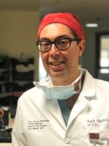 John Oghalai, MD, is a Professor and Department Chair of Otolaryngology-Head and Neck Surgery at USC. He has followed a clinician-scientist pathway, both caring for patients and performing research in the lab. His clinical areas of focus are acoustic neuroma (vestibular schwannoma) surgery, chronic ear disease, and cochlear implantation. His NIH-funded lab seeks to better understand the fundamental changes within the inner ear that underlie progressive hearing loss and to develop novel techniques to treat this problem before it worsens.
Prior to joining USC, he was an Instructor at UCSF from 2001-2003 where he did fellowship training in Neurotology and Skull Base Surgery. He joined the faculty at Baylor College of Medicine in 2003 as an Assistant Professor and was promoted to Associate Professor in 2009. In 2010, he moved to Stanford University as an Associate Professor, and was promoted to Professor in 2015. In 2017, he took his current position at USC.
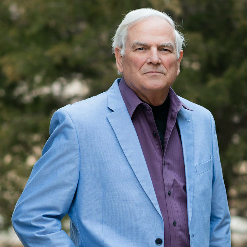 Robert M. Traynor, Ed.D., is a hearing industry consultant, trainer, professor, conference speaker, practice manager and author. He has decades of experience teaching courses and training clinicians within the field of audiology with specific emphasis in hearing and tinnitus rehabilitation. He serves as Adjunct Faculty in Audiology at the University of Florida, University of Northern Colorado, University of Colorado and The University of Arkansas for Medical Sciences.