
By Melissa A. Papesh and Frederick J. Gallun
Difficulty understanding speech in noisy environments is one of the most common complaints motivating patients to seek care from audiologists. Most often, this complaint is associated with some degree of hearing impairment, advancing age, or a combination of both. But how is an audiologist to respond when the patient is a young to middle-aged adult with no signs of peripheral hearing loss?
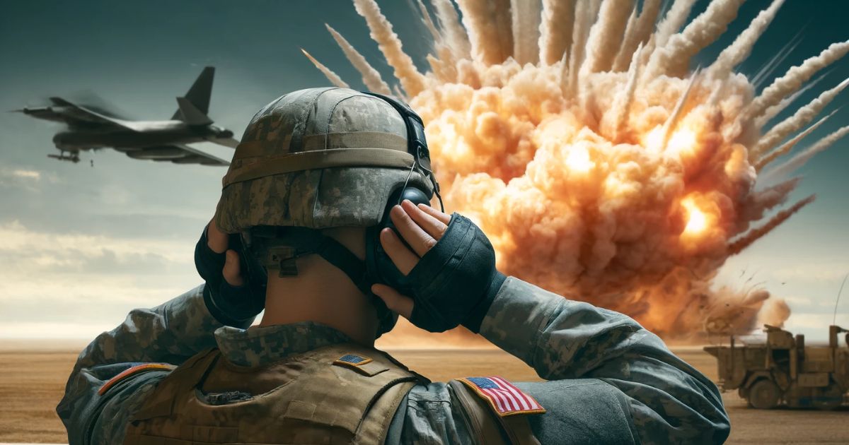
This situation is becoming more and more common in Department of Veterans Affairs (VA) and other audiology clinics across the country. On a recent survey of VA audiologists, 92% respondents reported seeing at least one patient per month with auditory complaints who was subsequently found to have normal or near normal hearing thresholds, and 39% of respondents indicated seeing an average of four or more such patients per month (Saunders and Echt, 2012). A common factor among many of these patients is combat experience including exposure to high-intensity blast waves. Military members involved in recent conflicts in Iraq and Afghanistan are being exposed to high-intensity explosives at unprecedented rates, largely due to our enemies’ heavy reliance on improvised explosive devices (IEDs).

Military members involved in recent conflicts in Iraq and Afghanistan have been exposed to high-intensity explosives at unprecedented rates
IEDs and other types of explosive weaponry account for approximately 75% of the casualties suffered by American soldiers during Operation Iraqi Freedom (OIF) and Operation Enduring Freedom (OEF) (Belmont et al., 2010). Though the toll on human life is significant, survival rates following injury are in fact vastly improved compared to previous military conflicts largely due to advancements in medical care in theater as well as improvements in armor and defensive gear (DOD, 2013). Overall, this situation has created thousands of Veterans living with the consequences of blast exposure while clinicians and researchers scramble to effectively diagnose and treat chronic symptoms of blast exposure.
With this in mind, the primary goal of the present work is to call attention to the auditory concerns of this ever growing population of Veterans, and to provide an overview of current research regarding the effects of blast exposure on the central auditory system.
Auditory Damage from Blast Exposure
When one first considers the auditory implications of blast exposure, initial thoughts are likely to focus on damage to peripheral portions of the auditory pathway such as tympanic membrane rupture, ossicular chain discontinuity, and potential damage to the inner ear (Cho et al., 2013; Fausti et al., 2009). Many peripheral injuries resolve spontaneously within a few months of injury (Cho et al., 2013; Mao et al., 2012) or respond favorably to surgical intervention. Lasting sensorineural hearing loss from blast-exposure is most often treated with the typical regime of hearing aids and assistive listening devices (Fausti et al., 2009).
More difficult to treat, however, are the large number of Veterans who complain of persistent hearing troubles in spite of normal hearing or mild hearing loss, implying a central source of dysfunction (Gallun et al., 2012b; Saunders and Echt, 2012). The likelihood of high-intensity blast waves impacting processing in the central auditory system seems high when one considers what is known about the interaction of blast waves with the brain. When a high-intensity explosive charge is detonated, it creates a region of extreme heat and pressure called a shock front which radiates outward at supersonic speeds. Exposure to the shock front results in what are called primary blast injuries as the wave of high pressure moves through bodily tissues.
When the shock front impacts the head, neural tissue is deformed due to stretching, compressing, and shearing forces as well as the impact of the brain against the skull (Cullis, 2001; Magnuson et al., 2012). These forces are likely to disrupt neural connections and damage blood vessels throughout the brain. Animal models indicate that the brain areas most likely to hemorrhage following blast exposure include the brainstem and midbrain (Knudson and Øen, 2003; Saljo et al., 2011), both of which comprise important components of the central auditory pathway. With regard to damaged neural connections, data from both animal models and patients with blast-induced TBI often indicate widespread diffuse axonal injury and resulting impaired connectivity between structures (Davenport et al., 2012; Garman et al., 2011). The disruption of axonal white matter following blast exposure is particularly prevalent in interhemispheric and intrahemispheric structures (Bauman et al., 2009; Lu et al., 2012) and between the cortical and subcortical structures (Davenport et al., 2012; Lu et al., 2012). In addition to damage to axons, damage to grey matter is also common.
Blast exposure has been associated with reduced volume measured in the hippocampus (Lu et al., 2012; Sajja et al., 2014) and in periventricular portions of the brainstem and midbrain (de Lanerolle et al., 2011; Panzer et al. 2012; Sarkar et al., 2011) including the inferior colliculus and medial geniculate body (Mao et al., 2012).
In addition to primary blast injuries, blast exposure may result in secondary blast injuries caused by bodily impact with shrapnel and other objects propelled by the explosion, tertiary blast injuries incurred when the individual is thrown to the ground or into other stationary objects, and/or quaternary injuries which encompass any additional blast-related damage such as burns (Wightman and Gladish, 2001). These latter types of blast injury generally result in overt physical damage for which a victim is likely to seek medical care. This is in contrast to primary blast injuries alone which often result in essentially invisible damage. Within the milieu of active combat, many service members are reluctant to report blast exposure to their superiors in the absence of physical damage, thus putting themselves at risk of sustaining additional injuries before their brains have properly healed. Such conditions may significantly increase the risk of permanent neurological damage (Mori et al., 2006; Cantu 1998; Saunders and Harbaugh, 1984). The propensity for sustaining multiple blast injuries marks a potentially important difference between blast exposure in Veterans and in the civilian population.
Veterans at Greater Risk
While military members are frequently exposed to multiple high-intensity blasts throughout their careers, the rare instances of blast exposure in civilian populations are usually isolated incidents. Data from other types of closed-head injuries such as sports-related concussions are now showing that multiple head traumas, even when diagnosed as mild, generally compound chronic neurological deficits (McKee et al., 2010; Mori et al., 2006). Thus, logic would dictate that Veterans are at greater risk for chronic impairments.
Though difficulty understanding speech in noisy environments is one of the most frequent hearing-related complaints of blast-exposed Veterans, other auditory complaints are also common. At the VA Medical Center in Tampa, Florida, which is home to one of the nation’s top five facilities designed to provide intensive rehabilitative care to Veterans and military members with traumatic brain injuries, audiologists have compiled a list of common auditory complaints of blast-exposed Veterans. These include difficulty understanding speech in noisy or reverberant environments, poor comprehension of rapidly spoken speech, difficulty following long conversations and recalling multiple spoken instructions, and the need for more time to process spoken information, among others. The majority of these Veterans are young and are often found to have normal or nearly normal hearing thresholds.
Studying Effects of Blast Exposure
The increasing number of such reports helped to initiate one of the few studies specifically designed to examine the effects of blast exposure on central auditory processing. This study was a collaborative effort between Walter Reed National Military Medical Center in Bethesda, Maryland and the National Center for Rehabilitative Auditory Research (NCRAR) located in the Portland, Oregon VA Medical Center. Study participants included military members exposed to at least one high intensity blast within the past year and who had no more than mild hearing loss at the time of testing.
Participants were predominately male with an average age of 32.8 years. While performance on a standard test of word recognition in quiet revealed no difference between the blast-exposed and non-blast-exposed age-matched control participants, blast-exposed participants displayed significantly poorer speech comprehension in noise as measured via the QuickSIN test (Gallun et al., 2012b). In addition, results indicated that blast-exposed participants were significantly more likely to perform poorly on two or more behavioral tests of central auditory processing compared to control participants (Gallun et al., 2012b).
Though performance across the test battery varied among individuals, tests on which blast-exposed participants were most likely to perform poorly included the Staggered Spondaic Words test (Katz et al, 1963), the Gaps in Noise test (Musiek et al., 2005), and the Masking Level Difference test (Wilson et al., 2003). The Staggered Spondaic Words test requires functional integrity of the temporal and frontal cortex and corpus callosum necessary for the processing of binaural competing speech (Katz and Smith, 1991). The Gaps in Noise test, administered monaurally, assesses temporal resolution and is sensitive to lesions throughout the central auditory system including the brainstem and the cerebrum (Musiek et al., 2005). The Masking Level Difference test assesses the binaural encoding of stimulus envelopes which is primarily performed by the brainstem (Noffsinger et al., 1982; Colburn, 1977). Hence, behavioral results indicate that auditory deficits from blast exposure may arise from damage to the auditory brainstem and/or cerebrum as well as non-auditory-specific structures such as the corpus callosum and frontal cortex.
Overall, the widespread nature of behavioral effects is congruent with knowledge of the potential for diffuse neural injuries from blast exposure.
In addition to behavioral tests of central auditory processing, two evoked potential measures were assessed including auditory brainstem responses and P300s. No significant group differences were found for either the latencies or amplitudes of ABR peaks indicating that brainstem encoding of acoustic onsets was intact following blast exposure. The same was not true for P300 measures in which 500 Hz tones served as the standard stimulus and 1000 Hz tones comprised the oddball stimulus. Responses to oddball stimuli clearly revealed a significant increase in latency and decrease in amplitude of P300 peaks in blast-exposed Veterans compared to control participants (Gallun et al., 2012b). Hence, while blast-exposed participants could perceptually distinguish between the pitch of the standard and oddball stimuli (as evidenced by their accurate counting of the oddball stimuli), the electrophysiological evidence revealed that their brains where slower and less robust at making this discrimination than non-blast exposed participants. This finding is in line with previous accounts of diminished and prolonged P300 potentials in patients with traumatic brain injury (TBI) from other causes (Lew et al., 2004; Folmer et al., 2011). Overall, the electrophysiological and behavioral test results of the study by Gallun et al. represent an important step in corroborating the subjective auditory concerns of blast-exposed Veterans.
Another important factor gleaned from the study by Gallun and colleagues (2012b) was that diagnosis of traumatic brain injury (TBI) was not a good predictor of participant performance on the test battery. Blast-exposed individuals with a medical diagnosis of mild TBI did not perform significantly poorer than their blast-exposed counterparts with no TBI diagnosis. Mild TBIs, commonly known as concussions, are inherently difficult to diagnose because they rarely produce damage visible with standard neuroimaging techniques (Ruff and Jurica, 1999). Therefore, diagnostic criteria for mild TBI typically rely upon subjective metrics most applicable to the immediate effects of the injury such as the patient’s report of loss of consciousness, altered mental state, and post-traumatic amnesia (Hoge et al., 2008; Ruff and Jurica, 1999). The similarity in performance between blast-exposed individuals with and without a TBI diagnosis (Gallun et al., 2012b) suggests that such metrics may not be sensitive to neural injury affecting functions such as auditory processing. Thus, clinicians should not rely upon a diagnosis of TBI alone when drawing conclusions about the potential for auditory effects from reported blast exposure in their patients. Rather, a better diagnostic picture may be gained by interviewing patients as to their military service history and proximity to high-intensity blast events.
The study by Gallun and colleagues (2012b) was one of the first to specifically target central auditory processing deficits in blast-exposed Veterans, and it provided compelling evidence that blast exposure can lead to measureable dysfunction even after a year of recovery. The question is now whether such auditory consequences persist, resolve, or worsen over the course of time. The critical need for such information was highlighted in a recent Institute of Medicine (IOM) report (IOM, 2014), generated in response to the Department of Veterans Affairs’ (VA) request for a comprehensive review of the scientific literature on the long-term health consequences of blast exposure. With regard to auditory function, the report found insufficient evidence of an association between blast exposure and central auditory processing dysfunction. Reasons cited included a distinct paucity of studies exploring central auditory consequences in this population, lack of standardized clinical test batteries for central auditory processing dysfunction, lack of information regarding the sites of injury and underlying mechanisms responsible for specific symptoms, and lack of knowledge regarding the effects of confounding factors such as medication use, cognitive and multisensory impairments, and comorbid conditions such as post-traumatic stress disorder (PTSD) and TBI from non-blast sources. This finding by the IOM committee emphasizes the need both for additional research on the auditory consequences of blast exposure as well as a consensus on the evaluation and diagnosis of central auditory processing disorders.
Currently, Gallun and colleagues at the NCRAR are addressing the lack of information on the chronic central auditory consequences of blast exposure by assessing central auditory effects in patients with blast exposure incurred within the past ten years. Preliminary evidence indicates a pattern strikingly similar to that found in the previous study both with regard to behavioral and electrophysiological test measures. In addition, new data regarding spatial release from masking tests indicate that blast-exposed Veterans often benefit less from spatial cues in order to improve speech detection. This finding adds another crucial piece of evidence which may explain why poor understanding of speech in noisy environments is such a common complaint in this population, and also implies that FM systems or low-gain directional hearing aids may be good options for rehabilitation (Gallun et al., 2012a; Fausti et al., 2009).
Blast Exposure and Lasting Auditory Impact
In summary, current research indicates that even mild cases of blast exposure can cause long lasting auditory consequences which may not be predicted by pure tone audiograms or recognition of speech in quiet. Considerable numbers of blast-exposed Veterans report life-altering auditory concerns from blast exposure in spite of thresholds indicating normal hearing. Thus, in cases where reported hearing difficulties are not explained by hearing loss or aging, audiologists should inquire about military service, blast exposure, and head injury.
Performance on tests of central auditory function indicates that auditory deficits in this population often include poorer temporal resolution, binaural processing, and auditory cognition (Gallun et al., 2012b).
While more research is needed to determine the most effective treatment options for this population, audiologists might consider devices which improve the signal-to-noise ratio in complex listening environments as well as encouraging patients to be proactive in modifying their acoustic environments to improve their listening experience when possible. Above all, it is critical that audiologists validate the concerns of this laudable patient population.
Melissa A. Papesh, Au.D., Ph.D., recently received a dual Ph.D. in Hearing Science and Neuroscience from Indiana University. She is also a licensed audiologist, a member of the American Speech-Language and Hearing Association (ASHA), and a Fellow of the American Academy of Audiology (AAA). Dr. Papesh is currently an Advanced Research Fellow in Polytrauma/TBI at the National Center for Rehabilitative Auditory Research in Portland, Oregon, awarded through the VA’s Office of Academic Affiliations. Her current research combines electrophysiological and behavioral test measures to probe sensory deficits associated with blast exposure and other types of head trauma.
Frederick J. Gallun, Ph.D. is a researcher at the National Center for Rehabilitative Auditory Research, and Associate Professor in Otolaryngology and the Neuroscience Graduate Program at Oregon Health and Science University. He received his degree in Cognitive Psychology from UC Berkeley and completed an NIH-funded postdoctoral fellowship at Boston University. His laboratory and research collaborations are funded by three NIH grants and three VA Merit Awards. The work focuses on the impacts of aging, hearing loss, and brain injury on the ability to parse the auditory scene, with an emphasis on spatial hearing and the processing of temporal information.
References:
- Bauman, R.A., Ling, G., Tong, L., Januszkiewicz, A., Agoston, D., Delanerolle, N., Kim, Y., Ritzel, D., et al. (2009). An introductory characterization of a combat-casualty-care relevant swine model of closed head injury resulting from exposure to explosive blast. Journal of Neurotrauma, 26(6), 841-860.
- Belmont, P.J., Schoenfeld, A.J., & Goodman, G. (2010). Epidemiology of combat wounds in Operation Iraqi Freedom and Operation Enduring Freedom: Orthopaedic burden of disease. Journal of Surgical Orthopaedic Advances, 19(1), 2-7.
- Cantu, R.C. (1998). Second-impact syndrome. Clinics in sports medicine, 17(1), 37-44.
- Colburn, H.S. (1977). Theory of binaural interaction based on auditory-nerve data. I. Detection of tones in noise. Journal of the Acoustical Society of America, 61, 525–533.
- Cullis, I.G. (2001). Blast waves and how they interact with structures. Journal of the Royal Army Medical Corps, 147(1), 16-26.
- Davenport, N.D., Lim, K.O., Armstrong, M.T., & Sponheim, S.R. (2012). Diffuse and spatially variable white matter disruptions are associated with blast-related mild traumatic brain injury. Neuroimage, 59(3), 2017-2024.
- de Lanerolle, N.C., Bandak, F., Kang, D., Li, A.Y., Du, F., Swauger, P., Parks, S., Ling, G., & Kim, J.H. (2011). Characteristics of an explosive blast-induced brain injury in an experimental model. Journal of Neuropathology & Experimental Neurology, 70(11), 1046-1057.
- Department of Defense (DOD). (2013). Principle Wars in which the United States Participated: U.S. Military Personnel Serving and Casualties. https://www.dmdc.osd.mil/dcas/pages/report_principal_wars.xhtml (accessed September 5, 2013).
- Fausti, S. A., Wilmington, D. J., Gallun, F. J., Myers, P. J., & Henry, J. A. (2009). Auditory and vestibular dysfunction associated with blast-related traumatic brain injury. Journal of Rehabilitation Research & Development, 46(6), 797-810.
- Folmer, R. L., Billings, C. J., Diedesch-Rouse, A. C., Gallun, F. J., & Lew, H. L. (2011). Electrophysiological assessments of cognition and sensory processing in TBI: applications for diagnosis, prognosis and rehabilitation. International Journal of Psychophysiology, 82(1), 4-15.
- Gallun, F. J., Lewis, M. S., Folmer, R. L., Diedesch, A. C., Kubli, L. R., McDermott, D. J., … & Leek, M. R. (2012a). Implications of blast exposure for central auditory function: A review. Journal of Rehabilitation Research & Development, 49(7), 1059-74.
- Gallun, F.J., Diedesch, A.C., Kubli, L.R., Walden, T.C., Folmer, R.L., Lewis, M.S., McDermott, D.J., Fausti, S.A., & Leek, M.R. (2012b). Performance on tests of central auditory processing by individuals exposed to high-intensity blasts. Journal of Rehabilitation Research and Development, 49(7), 1005–1024.
- Garman, R.H., Jenkins, L.W., Switzer III, R.C., Bauman, R.A., Tong, L.C., Swauger, P.V., Parks, S.A., Ritzel, D.V. et al. (2011). Blast exposure in rats with body shielding is characterized primarily by diffuse axonal injury. Journal of Neurotrauma, 28(6), 947-959.
- Hoge, C. W., McGurk, D., Thomas, J. L., Cox, A. L., Engel, C. C., & Castro, C. A. (2008). Mild traumatic brain injury in US soldiers returning from Iraq. New England Journal of Medicine, 358(5), 453-463.
- IOM (Institute of Medicine) (2014). Gulf War and health, volume 9: Long-term effects of blast exposures. Washington, DC: National Academies Press.
- Katz, J., Basil, R. A., & Smith, J. M. (1963). A staggered spondaic word test for detecting central auditory lesions. The Annals of otology, rhinology, and laryngology, 72, 908-917.
- Katz, J. & Smith, P.S. (1991). The staggered spondaic word test. Annals of the New York Academy of Sciences, 620(1), 233-251.
- Knudsen, S.K. & Øen, E.O. (2003). Blast-induced neurotrauma in whales. Neurosci Res, 46(3), 377-386.
- Lew, H. L., Lee, E. H., Pan, S. S., & Date, E. S. (2004). Electrophysiologic abnormalities of auditory and visual information processing in patients with traumatic brain injury. American journal of physical medicine & rehabilitation, 83(6), 428-433.
- Lu, J., Ng, K.C., Ling, G., Wu, J., Poon, D.J.F., Kan, E.M., Tan, M.H., Wu, Y.J. et al. (2012). Effect of blast exposure on the brain structure and cognition in Macaca fascicularis. Journal of Neurotrauma, 29(7), 1434-1454.
- Magnuson, J., Leonessa, F., & Ling, G.S. (2012). Neuropathology of explosive blast traumatic brain injury. Current Neurology & Neuroscience Reports, 12(5), 570-579.
- Mao, J. C., Pace, E., Pierozynski, P., Kou, Z., Shen, Y., VandeVord, P., … & Zhang, J. (2012). Blast-induced tinnitus and hearing loss in rats: behavioral and imaging assays. Journal of neurotrauma, 29(2), 430-444.
- McKee, A.C., Cantu, R.C., Nowinski, C.J., Hedley-Whyte, E.T., Gavett, B.E., Budson, A.E., … & Stern, R. A. (2009). Chronic traumatic encephalopathy in athletes: progressive tauopathy following repetitive head injury. Journal of neuropathology and experimental neurology, 68(7), 709.
- Mori T, Katayama Y, & Kawamata T. (2006). Acute hemispheric swelling associated with thin subdural hematomas: pathophysiology of repetitive head injury in sports. In Brain Edema XIII. 40-43. Springer Vienna.
- Musiek FE, Shinn JB, Jirsa R, Bamiou DE, Baran JA, Zaida E. (2005). GIN (Gaps-In-Noise) test performance in subjects with confirmed central auditory nervous system involvement. Ear and Hearing, 26(6), 608–18.
- Noffsinger, D., Martinez, C.D., & Schaefer, A.B. (1982). Auditory brainstem responses and masking level differences from persons with brainstem lesion. Scandinavian Audiology Supplementum, 15, 81–93.
- Panzer, M.B., Myers, B.S., Capehart, B.P., & Bass, C.R. (2012). Development of a finite element model for blast brain injury and the effects of CSF cavitation. Annals of Biomedical Engineering, 40(7), 1530-1544.
- Ruff, R.M. & Jurica, P. (1999). In search of a unified definition for mild traumatic brain injury. Brain Injury, 13(12), 943-952.
- Sajja, V.S.S.S., Ereifej, E.S., & VandeVord, P.J. (2014). Hippocampal vulnerability and subacute response following varied blast magnitudes. Neuroscience Letters, 570, 33-37.
- Säljö, A., Mayorga, M., Bolouri, H., Svensson, B., & Hamberger, A. (2011). Mechanisms and pathophysiology of the low-level blast brain injury in animal models. Neuroimage, 54, S83-S88.
- Sarkar, A, Yashwanth, BL, & Sarkar, S. (2011). Analysis of Blast Induced Intracranial Pressure Dynamics in Cerebrospinal Fluid Leading to Traumatic Brain Injury. International Journal of Emerging Multidisciplinary Fluid Sciences, 3(2), 135-144.
- Saunders, G.H. & Echt, K.V. (2012). Blast exposure and dual sensory impairment: An evidence review and integrated rehabilitation approach. Journal of Rehabilitation Research and Development, 49(7), 1043–1058.
- Saunders, R.L. & Harbaugh, R.E. (1984). The second impact in catastrophic contact-sports head trauma. Journal of the American Medical Association, 252, 538-9.
- Wightman, J.M. & Gladish, S.L. (2001). Explosions and blast injuries. Annals of Emergency Medicine, 37(6), 664-678.
- Wilson, R.H., Moncrieff, D.W., Townsend, E.A., Pillion, A.L. (2003). Development of a 500-Hz masking-level difference protocol for clinic use. Journal of the American Academy of Audiology, 14(1), 1–8.


Do you have any information bout the onset of stuttering after blast exposure?