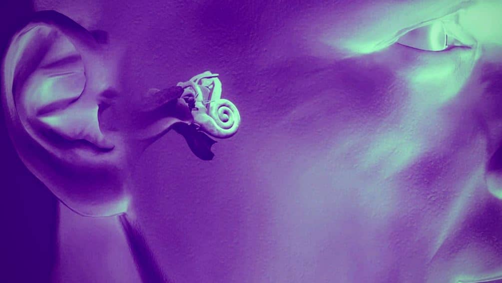
Third mobile window syndromes (TMWS) are a relatively recent discovery but are an increasingly recognized pathology in otology. The hearing function of the human ear works by the pinna and the external auditory canal funneling sound to the eardrum. The eardrum vibrates once stimulated with the sound waves and moves the middle ear bones called the ossicles. The ossicles then work like a piston, pushing in on the fluid-filled inner ear cochlea via the oval window, and the round window acts as a contained pressure release valve of sorts, distending with movement of the oval window.
This fluid disturbance in the inner ear is then captured by the cochlear hair cells to be relayed to the cochlear nerve, which carries the information to the brain for the perception of sound.

A TMWS is a condition where a third mobile area in the inner ear exists, most often due to abnormalities in the bone surrounding the membranous inner ear structures. This alters the fluid dynamics of the system and can result in a multitude of seemingly unusual symptoms including but not limited to:
- Dizziness provoked by loud sounds (Tullio phenomenon)
- Dizziness associated with physical strain (Hennebert sign)
- One’s own voice sounding unusual (autophony)
- Pulsatile tinnitus
- Hearing loss
- Hearing internally generated sounds such as eye movements, chewing, or footsteps.
Due to the unusual nature of some of the symptoms associated with TMWS, many of those suffering from these conditions are misdiagnosed or endure a prolonged period of time to reach the correct diagnosis.
Semicircular Canal Dehiscence
The human inner ear consists of three angular sensors known as the superior, lateral and posterior semicircular canals. These canals serve a primary role of visual stability during head rotation through the vestibular ocular reflex. An abnormal opening or dehiscence along one of these canals causes the canal to be stimulated with sound, physical strain, or change in middle ear pressure. When the inner ears receive conflicting signals, this leads to an abnormal eye movement called nystagmus, which is perceived as a sensation of vertigo or dizziness.
The most well studied TMWS is the condition of superior semicircular canal dehiscence syndrome (SCDS). Dr. Loyd Minor at Johns Hopkins University first described SCDS in 1998.
SCDS is the result of a thinning or break in the bone that separates the superior semicircular canal from the brain. The condition is felt to be congenital in most cases but can also be acquired through head injury. The prevalence of SCDS has been shown to be between 2-10% on CT scans of the temporal bone. The dehiscence can often be surgically repaired with good outcomes for those individuals with debilitating symptoms.
Dehiscence of the posterior (PCDS) and lateral canal (LCDS) is much less common affecting 1.2% and 0.4% of temporal bones respectively.

Image courtesy earsite.com with modifications
Dehiscence of the posterior semicircular canal has been shown to be most often associated with a high riding jugular bulb, where the jugular vein is essentially in too close of proximity with the inner ear structures. A dehiscence of the lateral semicircular canal is typically associated with chronic middle ear infections that lead to a mass of infection called a cholesteatoma, which erodes into the semicircular canals.
Cochlear Facial Dehiscence
Cochlear facial dehiscence refers to a thinning or break in the boney structure separating the cochlea from the facial nerve.
This condition is an even more recent discovery that was first described in 2014. Less is known about this type of dehiscence but a recent study found cochlear facial dehiscence in around 0.6% of temporal bones. This same study indicated that the condition is likely developmental and associated with being older, a female, or caucasian.
Cochlear Carotid Dehiscence and Cochlear Internal Auditory Canal Dehiscence
A cochlear carotid dehiscence is a thinning or break in the bone that separates the carotid artery from the cochlea.
A cochlear IAC dehiscence is a thinning or break in the bone that separates the cochlea from the internal auditory canal where the hearing and balance nerve travels to the brain.
Both of these conditions are thought to be quite rare and at the time of writing, I am unaware of any large-scale temporal bone studies investigating these pathologies. Due to the lack of knowledge on these conditions, there is little information on at risk populations or prevalence data. Only a handful of case reports exist describing these conditions. Links to a few of these case reports can be found below:
Carotid-artery cochlear dehiscence
Cochlear-Internal Canal and Cochlear-Facial Dehiscence: A Novel Entity
Carotid artery-cochlear dehiscence: a review

(A) Cochlear-carotid dehiscence (Modugno et al.,3 Kim and Wilson,4 Neyt et al.,5 Lund and Palacios6). (B) Cochlear-internal auditory canal dehiscence (Karlberg et al.,7 Manzari and Scagnelli8). (C) Cochlear-facial dehiscence (reported in this study). (D) Cochlear-jugular dehiscence (not yet described). (Image from Cochlear-Facial Dehiscence- A Newly Described Entity, Blake et al. 2014)
Enlarged Vestibular Aqueduct (EVA)
The endolymphatic sac regulates inner ear fluid and is responsible for the immune response of the inner ear. The vestibular aqueduct is the fluid pathway that connects the endolymphatic sac to the other parts of the inner ear system.
Enlarged vestibular aqueduct (EVA) is a congenital condition where the aqueduct is too large which can result in hearing loss and vestibular symptoms.
EVA is one of the most common inner ear malformations in children with hearing loss. It has been shown that 5-15% of children with sensorineural hearing loss have EVA. There are currently very few studies describing the specific dizziness symptoms associated with EVA. We do know that those with EVA often report vestibular symptoms and have evidence of vestibular dysfunction on testing. It seems possible and it has been reported that an enlarged vestibular aqueduct could potentially create a third mobile window effect on the ear.
Perilymph Fistula
A perilymph fistula refers to a hole or fistula that allows inner ear perilymph to leak out. This leak is thought to most commonly occur at the level of the round or oval window, which allows the perilymph to leak into the middle ear space. This is a somewhat controversial diagnosis, largely because it is difficult to assess for this condition.
Perilymph fistula is felt to have an incidence of 1.5/100,000. Some potential causes for the condition include rapid changes in barometric pressure, trauma including temporal bone fracture, as well as postsurgical complications.
Summary
TMWS are a recent discovery that can cause multiple dizziness and aural symptoms. The conditions described are simply the TMWSs that we are currently aware of and it is likely that others will be made known in the future. Those with TMWS are often misdiagnosed or do not receive a diagnosis at all.
Increased awareness of these conditions is the first step to ensure correct and timely diagnoses for those living with a TMWS.


