A vestibular evoked myogenic potential, or VEMP, is a measure of vestibular function that only recently is seeing widespread clinical use. A VEMP is a measurement of a change in muscle activity in response to stimulating the vestibular system. These measures allows us to intuit the function of specific end organs of the inner ear, predominantly reflecting the function of the otolith organs (saccule and utricle).
These measures are typically completed with either acoustic or bone conducted clicks or tone bursts. Essentially, these acoustic or bone-conduction stimuli are applied either to the ear canal or skull to shake the otolith organs. The utricle and saccule have separate projections to different parts of the body. By recording in different locations on the body, we can determine how each of these end organs are functioning.
The two most commonly completed VEMP measures are the cervical and ocular VEMP:
- The cVEMP is thought to predominantly reflect the function of the saccule and/or inferior branch of the vestibular nerve
- The oVEMP is thought to predominantly reflect the function of the utricle and/or superior branch of the vestibular nerve
These responses do rely on an intact central nervous system and brainstem structures in order to transmit these responses to the respective muscle groups that the responses are recorded from. More can be found on the details of the most commonly utilized VEMPs and how they are utilize clinically here and here.
Additional studies have investigated other potential sites for recording VEMP responses. Some studies have shown VEMP responses that are capable of being recorded from the soleus, biceps, gastrocnemius, triceps, and deltoids. These additional recording sites are not typically used in a clinical setting but may have the capability to provide additional information in certain patient populations such as this with spinal cord injury, progressive neuro muscular disease, or even in cases of stroke. We do not currently know all of the potential uses of these different VEMP measures.
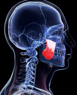
The Masseter VEMP (mVEMP)
Recently there has been investigation into the recording of a VEMP response from the masseter muscle of the jaw. At the time of writing this, this measure is thought to reflect the function of both the saccule and cochlea. There are reports of an initial vestibular/saccule component with latencies of p11/n15, overlapped with a cochlear component with latencies of p16/n21.
The response is bilateral and can be measured from either masseter, regardless of which ear is stimulated.

Masseter VEMP could be a useful tool to assess the function of the saccule
The response can be elicited with stimulus parameters similar to what are used for a cVEMP. A 500 Hz tone burst stimulus that is presented at around 100 dB nHL seems to be a reliable means of generating an mVEMP response. The mVEMP appears to scale with higher degrees of EMG, as well as higher stimulus presentation levels, just like most other VEMP measures. I was able to record my own mVEMP response on our Biologic AEP system with a 500 Hz tone burst, presented at 95 dB nHL. Additional information on stimulus recording parameters for mVEMP can be found here and here.
More data needs to be gathered on the mVEMP in order to gain consensus on the most appropriate parameters for stimulation, ideal EMG targets, as well as the best electrode montage. Also, given the cochlear contributions, it would seem prudent to have normative data for individuals with normal hearing and for those with sensorineural hearing loss.
Clinical Applications
The masseter VEMP could be a useful tool to assess the function of the saccule in patients that are unable to undergo cVEMP testing. Additionally, the mVEMP could be useful for certain patient populations with neurological disorders impacting the brainstem.
At this time, the mVEMP needs to be investigated more thoroughly prior to widespread clinical use, but it does appear to be an additional VEMP measure that could be included in a vestibular test battery.


