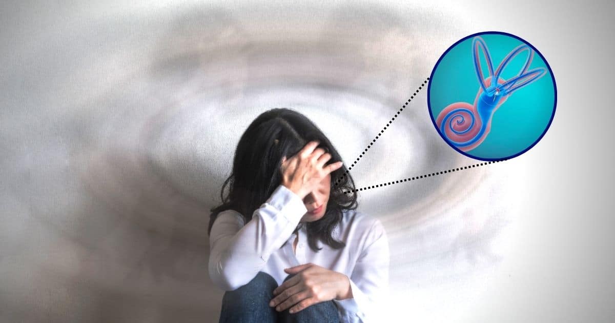
Ménière’s disease, a rare and debilitating condition that causes severe balance disorders and other distressing symptoms like nausea, spinning dizziness (vertigo), tinnitus, and hearing loss, has long puzzled medical experts.
Treatments between the intense episodes of vertigo that are characteristic of the condition vary, and may include dietary changes, diuretics, and steroids. For more severe cases, aggressive treatments, such as injecting antibiotics into the ear or even surgical procedures, can provide relief but come with risks of permanent vestibular (balance) deficits.
Ménière’s Disease and Inner Ear Volume
In an exciting breakthrough, a successful collaboration between the Karl Landsteiner University of Health Sciences (KL Krems) in Austria and renowned institutions Harvard Medical School and Johns Hopkins University in the United States has shed new light on the disease’s underlying processes.
The research involved detailed 3D analysis of the inner ear, providing valuable insights into volume changes in specific structures associated with Ménière’s disease.
The team, led by Dr. Béla Büki from Krems University Hospital, compared the inner ears of nine Ménière’s patients with those of ten healthy individuals. By creating digital 3D models based on numerous anatomical slices, the team could measure the volumes of essential compartments in the inner ear, including the cochlear duct and the saccule and utricle Additionally, they analyzed the thickness of special membranes known as Reissner’s membrane and investigated the condition of the enigmatic “Bast’s valve.”
“Very often, the volume of the external cochlear duct as well as the one of the sacculus was enlarged in affected patients. We were able to clearly demonstrate this in the virtual 3D models.”
–Dr. Béla Büki
Researchers also found that the volume of the utricle increased, although less frequently than the other compartments.
Through the detailed analysis, the team measured the thickness of the membranes lining the compartments, revealing their mechanical resistance to the increased pressure of the inner ear fluids, known as endolymph. The membrane thickness correlated perfectly with the analyzed volumes of the compartments. Notably, the thicker Reissner’s membrane in the utricle of healthy individuals appeared to prevent volume expansion when endolymph pressure increased, potentially explaining why the utricle was less frequently dilated.
Bast’s Valve: Inner Ear Mystery for Nearly 100 Years
However, the utricle’s dilation in some affected individuals posed a question. Further investigation into the mysterious Bast’s valve, located at the entrance of the utricle, provided answers.
“In all cases of Ménière’s disease where the utricle was swollen, the researchers observed that the Bast’s valve was open or the surrounding membrane had ruptured. This finding suggested a pressure-regulating function for the valve, which remains a topic of scientific intrigue nearly a century after its discovery.”
The successful collaboration between KL Krems, Harvard Medical School, and Johns Hopkins University marks a significant step forward in understanding Ménière’s disease, contributing valuable knowledge to improve treatment outcomes for patients. This research aligns with KL Krems’ commitment to creating genuine clinical advancements that benefit those living with serious medical conditions.
Reference:
- B. Büki, B. K. Ward & F. Santos. Differential Volume Increase of Endolymphatic Compartments in Ménière’s Disease Is Inversely Associated With Membrane Thickness. Otol Neurotol. 2023 Jul 18. DOI:10.1097/MAO.0000000000003960
Source: KL Krems