
A groundbreaking study recently published in Audiology and Neurotology presents a novel application of synchrotron radiation phase-contrast imaging (SR-PCI) to examine cochlear otosclerosis.
This advanced imaging technique has enabled researchers to visualize otosclerotic lesions in unprecedented detail, offering new insights into the pathology and potential treatment avenues for this common cause of acquired hearing loss.
Otosclerosis
Otosclerosis is a bone disorder affecting the labyrinthine capsule of the inner ear, leading to both conductive and sensorineural hearing loss. Despite its prevalence, particularly among Caucasians, the exact etiology of otosclerosis remains unclear, with factors such as genetic predisposition, viral infections, hormonal changes, and autoimmunity all under consideration.
Traditionally, treatment has primarily involved surgical interventions, such as stapedectomy and even cochlear implantation, with limited success using medical therapies like sodium fluoride and bisphosphonates.
Research Methodology
The study, led by researchers from Uppsala University in Sweden and Western University in Canada, utilized SR-PCI to analyze otosclerotic plaques in a temporal bone obtained from a cadaver. This is the first time SR-PCI has been used to examine such lesions, marking a significant advancement in the field.
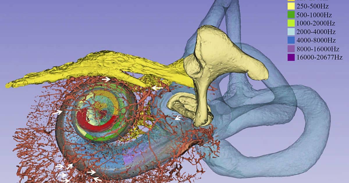
The technique allowed the researchers to create three-dimensional (3D) renderings of the otosclerotic lesions, providing detailed visualizations of their structure and vascular connections.
Images credit: Dina Giese, Helge Rask-Andersen, Hanif M. Ladak, Sumit Agrawal, Hao Li; Synchrotron Phase-Contrast Imaging and Cochlear Otosclerosis: A Case Report. Audiol Neurotol 2024; https://doi.org/10.1159/000539422
Two cochleae were analyzed: one from a patient with otosclerosis who had previously undergone a partial stapedectomy with a stapes wire prosthesis, and one from a healthy control. The samples were fixed in formaldehyde and imaged at the Canadian Light Source using the Biomedical Imaging and Therapy beamline. The resulting images were processed using 3D Slicer software to segment and render the lesions and surrounding structures.
Key Findings
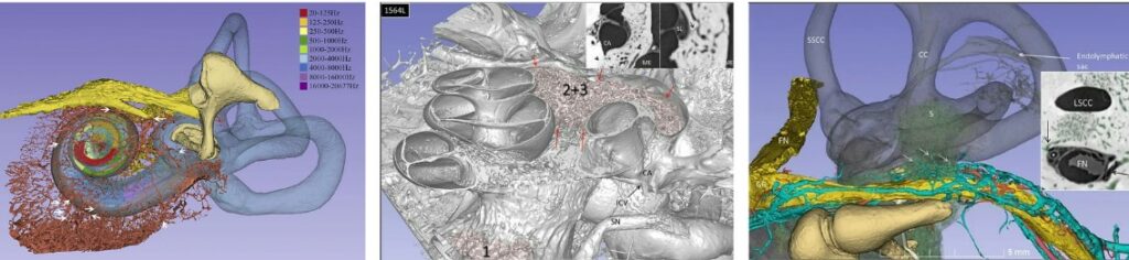
The study revealed several critical findings about the nature and impact of otosclerotic lesions. The lesions were found to be three-dimensionally distributed around the oval window, round window, and other regions of the cochlea. Notably, the study identified abnormal vascular connections between the otosclerotic foci and the cochlear blood supply. These vascular shunts and hypervascularization could lead to venous congestion, potentially explaining the sensorineural hearing loss observed in some patients.
One of the most significant findings was the involvement of the inferior cochlear vein (ICV) and cochlear aqueduct (CA) in the otosclerotic process. The ICV was significantly dilated in the otosclerotic ear, suggesting an overloading of the venous system. This vascular congestion could disrupt the microcirculation within the cochlea, leading to degeneration of sensory structures such as the organ of Corti and spiral ganglion cells.
Implications for Treatment
The detailed visualization of otosclerotic lesions provided by SR-PCI opens new possibilities for treatment. The researchers suggest that local pharmacotherapy targeting the middle ear could be a viable strategy to arrest the progression of sensorineural hearing loss in otosclerosis. By reducing angiogenesis and restoring normal bone metabolism, such treatments could alleviate the venous congestion and microcirculatory disturbances caused by the lesions.
Additionally, the study emphasizes the potential for SR-PCI to enhance the understanding of otosclerosis and guide the development of targeted therapies. The ability to visualize the intricate vascular connections and structural changes within the cochlea offers a powerful tool for both diagnosing and treating this complex disorder.
As research in this area continues, the findings from this study could help pave the way for improved diagnosis and treatment of otosclerosis, benefiting both clinical professionals and patients suffering from this challenging condition.
Citation:
- Dina GieseHelge Rask-AndersenHanif M. LadakSumit AgrawalHao Li; Synchrotron Phase-Contrast Imaging and Cochlear Otosclerosis: A Case Report. Audiol Neurotol 2024; https://doi.org/10.1159/000539422
Source: Audiology and Neurotology
*Featured image credit, Giese, et al.