A team from Western University has utilized the bright light of the Canadian Light Source (CLS) at the University of Saskatchewan to obtain highly detailed images of the intricate structures in the inner ear. These images have enabled them to pioneer customized programming strategies for hearing implants.
The cochlea, the spiral-shaped structure within the inner ear responsible for transmitting sound signals to the brain, is enclosed in the densest bone in the human body. This makes it challenging to study its anatomy and how implants interact with it using conventional techniques. However, synchrotron imaging has revolutionized this field by allowing scientists to visualize the cochlea in incredible detail, approximately at the scale of individual cells.
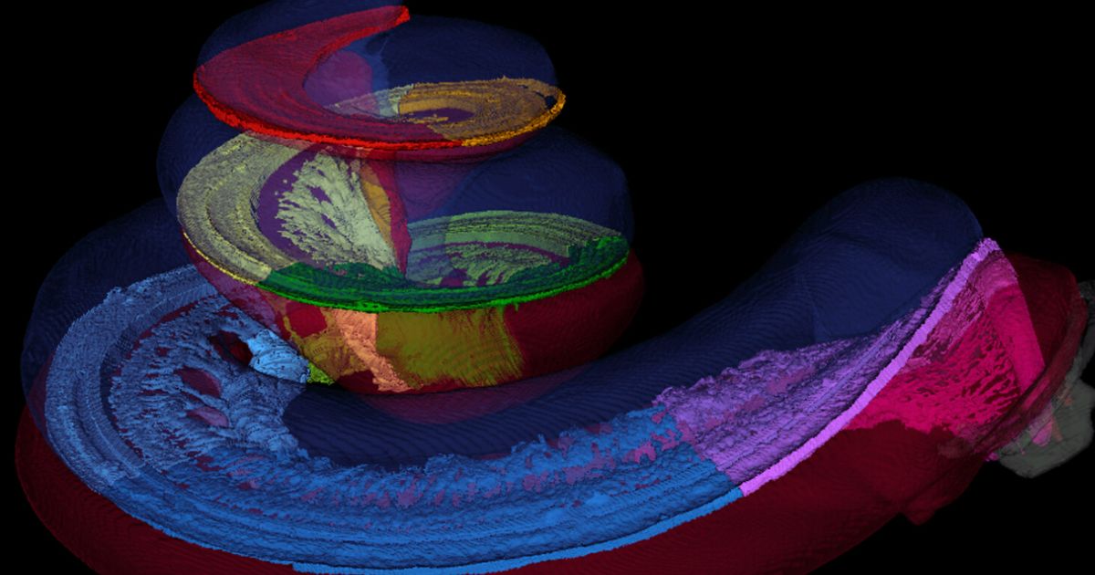
“We were able to obtain high-resolution data on the synchrotron, and then created beautiful three-dimensional images with our collaborators in Sweden”
–Dr. Sumit Agrawal from Western University
The team recently published the CLS-enabled mappings of 38 cochleae in the journal Laryngoscope. Agrawal states that this “gold standard data,” based on ultra-detailed imaging of the ear’s anatomy, answers many questions in the field.
Customized Maps to Improve Sound Quality for Cochlear Implants
These maps should significantly improve the sound quality of cochlear implants. As sound travels through the cochlea, different pitches land at different points in the structure for us to hear them. To tune the sound accurately, an implant needs to match these points for each individual patient’s anatomy. Without a map of the inner ear, cochlear implants can only be “one size fits all.”
“It would be like listening to an out-of-tune piano. What we’re doing now is actually mapping each of the electrodes to tune the piano for each individual patient”
By combining high-resolution imaging from the Bio-Medical Imaging and Therapy (BMIT) facility at the CLS with the team’s deep learning algorithms, researchers can now create customized maps that match the unique anatomy of each patient’s cochlea. The deep-learning algorithm was also partly trained on 3D images produced at the CLS.
The team has already begun applying this data to patients with cochlear implants and published a small pilot study using both customized and standard programming. The personally “tuned” implants showed promising results, providing the data needed to proceed with the randomized controlled trial currently underway at Western University and the University of North Carolina.
“The fact that we were able to extract the anatomy from the synchrotron, create a new equation, apply it to patients and improve their performance was really exciting for us,” says Agrawal.

A major grant from MED-EL, a leading hearing implant company, has helped guarantee the team can continue their ambitious synchrotron-enabled research program for many years to come. The $8.5-million donation, announced in late 2023 and matched by Western University, establishes two endowed research chairs devoted to this independent hearing research.
“Although we have already started applying this translational ‘bench-to-bedside’ research in patients, we are not going to stop using the synchrotron. There are so many other things we can look at, and the synchrotron allows us to see processes in real time,” says Agrawal. “This is just the start.”
Reference:
Micuda, Ashley, Hao Li, Helge Rask‐Andersen, Hanif M. Ladak, and Sumit K. Agrawal. “Morphologic Analysis of the Scala Tympani Using Synchrotron: Implications for Cochlear Implantation.” The Laryngoscope (2024). https://doi.org/10.1002/lary.31263
**Featured image: 3D color rendering of synchrotron radiation phase-contrast image of a cochlea, courtesy of Hao Li and Helge Rask-Andersen, Uppsala University Sweden
Source: CLS