
LOS ANGELES, CALIFORNIA — Researchers at the Keck School of Medicine of USC have adapted a low-cost imaging method originally developed for ophthalmology and used it to visualize tiny structures in the human inner ear.
Optical coherence tomography (OCT), a tool routinely used to diagnose and manage eye diseases, has now been modified to image the inner ear. A proof-of-concept study led by Keck School researchers found that OCT can measure fluid levels in the inner ear—an indicator that correlates with a patient’s degree of hearing loss. The findings were recently published in Science Translational Medicine.
“These findings are exciting because hearing loss can happen very suddenly, and we often don’t know why. OCT offers a way to explore the underlying cause and potentially guide treatment,” said senior author John Oghalai, MD, professor and chair in the Caruso Department of Otolaryngology – Head and Neck Surgery and the Leon J. Tiber and David S. Alpert Chair in Medicine at the Keck School of Medicine.
A New Way to Visualize Inner Ear Fluids
Sudden hearing loss—sometimes accompanied by vertigo—can occur in conditions such as Ménière’s disease, cochlear hydrops, and other inner ear disorders. A hallmark of these diseases is an imbalance of fluids in the inner ear, but accurately measuring these fluid levels has long been a challenge. Current imaging technologies like MRI lack the resolution needed to support reliable diagnosis and treatment decisions.
*A mouse cochlea imaged using OCT. Video credit: Oghalai Lab
OCT offers a quicker, more accurate, and more cost-effective way to visualize inner ear fluids, hair cells, and other critical structures. With funding from USC and the National Institutes of Health, Oghalai, Brian Applegate, PhD (professor of otolaryngology, ophthalmology, biomedical engineering, and electrical and computer engineering), and their team tested OCT in 19 patients undergoing ear surgery.
The tool reliably detected fluid imbalances, which correlated with the severity of hearing loss. Though currently limited to use during surgery, the researchers aim to adapt it for clinical settings.
The latest findings build on prior work where the team used OCT to image the cochlea in awake animals for the first time. With continued development, the tool could help clinicians quickly identify the causes of hearing loss, guide appropriate treatments, and even support the creation of new therapies to restore hearing.
Toward Faster, More Personalized Hearing Care
OCT works by scanning tissue with light waves to create detailed 3D images—similar to how ultrasound uses sound waves. In this study, the Keck team imaged the inner ears of 19 surgical patients: six with normal inner ear function, four with Ménière’s disease, and nine with vestibular schwannoma. During surgery, the outer mastoid bone was temporarily removed, allowing researchers to use OCT to measure fluid compartments.
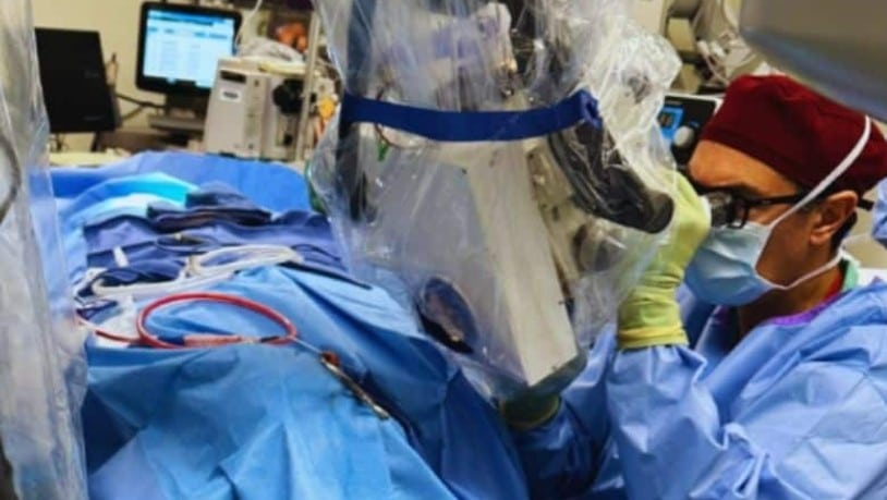
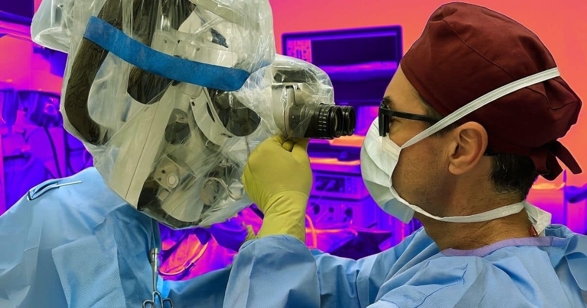
John Oghalai MD, is collecting data using Optical coherence tomography (OCT) while performing surgery. Credit: John Oghalai.
Patients with Ménière’s disease or vestibular schwannoma had higher levels of endolymph, a fluid associated with greater hearing loss. These findings suggest that measuring endolymph levels could help predict symptom severity.
“We’ve known for a long time that endolymph is related to hearing loss, but until now, measuring it in a living patient has been a major challenge”
—John Oghalai, MD
Beyond surgical applications, OCT may help clinicians distinguish between tumors and healthy tissue and avoid damage to delicate ear structures. To broaden its utility, the research team is developing a smaller, more affordable version of the device for use in operating rooms and eventually in outpatient clinics.
The team has received additional federal funding and a Nemirovsky Engineering and Medicine Opportunity (NEMO) Prize from USC to improve the software and imaging techniques needed for non-invasive, in-clinic use. Unlike MRI, OCT is fast, affordable, and suitable for repeated testing to monitor treatment response.
“Patient symptoms—sensations of hearing loss, pressure in the ear, or vertigo—can be affected by many unrelated factors such as mood, stress, nasal congestion and allergies. However, inner ear fluid levels reliably assess what is going on inside the inner ear,” said Oghalai. “We have lots of treatment options available, but it often takes many weeks to figure out what works for a given patient. This could be a faster way to help.”
If adapted for routine use outside the operating room, OCT could also support the development of new therapies for hearing loss. Gene therapies aimed at regenerating inner ear hair cells are currently being tested in clinical trials. OCT may provide a precise, real-time method for evaluating the effectiveness of these treatments.
“OCT helped researchers develop treatments for retinal diseases, including macular degeneration, so we’re hoping it can have a similar impact for hearing loss,” Oghalai said.
About this research
In addition to Oghalai and Applegate, the study’s other authors are Wihan Kim, Dorothy W. Pan, Bong Jik Kim, Zihan Yang, Marcela Moran Mojica, Joni K. Doherty and Seiji B. Shibata from the Caruso Department of Otolaryngology – Head and Neck Surgery, Keck School of Medicine of USC, University of Southern California.
This work was supported by the National Institute on Deafness and Other Communication Disorders of the National Institutes of Health [R01DC022232, R25DC019700]; the National Institute of Biomedical Imaging and Bioengineering of the National Institutes of Health [R01EB027113]; and University of Southern California Nemirovsky Engineering and Medicine Opportunity (NEMO) Prize.
Disclosure: John Oghalai and Brian Applegate are founders of AO technologies, with the goal of translating inner ear imaging technologies for clinical purposes.

Source: USC