 Ear Canal Dimensional Measurement Summary of Previous Five Posts
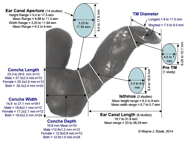
The previous five posts on the ear canal contained detailed published measurements and methods for making those measurements. This post takes those data and provides a summary illustration that shows the complete ear canal and highlights the locations of those dimensional measurements (Figure 1).
The data highlighted in black text is a summary of measurements from immediate previous posts on this site, and relates specifically to the ear canal. The text in blue provides information about features of the auricle (pinna), which is not part of the ear canal. The exception to this is that the TM (tympanic membrane) diameter is posted in blue, but is part of the ear canal. The remainder of data in blue text relates to the concha, an important area for accepting devices that are intended to be in-ear devices, but which mostly fit in the concha primarily. A future post will look at these measurements in greater detail, along with providing the references for the measurement data shown.

Figure 1. Dimensions of the ear canal superimposed on an ear impression of the complete human ear canal.
(Next week’s post will continue with the ear canal, and will focus on non-measurement features, such as the tissue lining, underlying structures, blood supply, innervation, etc.)


