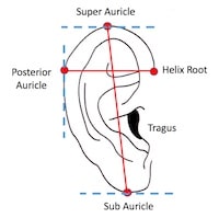
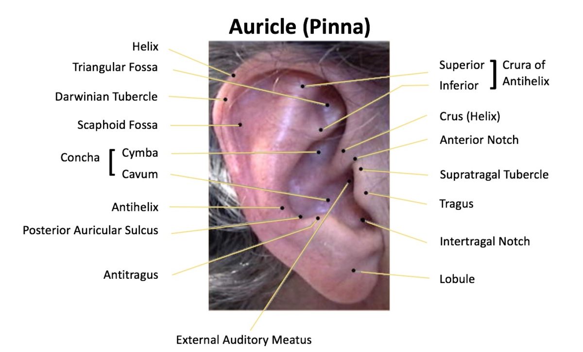
This post is a second in a series related to the anatomy of the human ear, specifically on the pinna, or auricle (Figure 1). Part 1 of this series started by identifying and defining the main external structural features of the auricle. This post concentrates on two primary dimensions of the auricle – length and width.
The anatomy of the external ear, also known as the auricle or pinna, is complex and remarkably inaccurately described by many authors. A previous post mentioned that even though ear anatomy is used differently by various disciplines (surgeons, dysmorphologists, anatomists, artists, forensics personnel, hearing aid dispensers, listening product engineers, patent writers etc.), it is incumbent that identification features and terminology be consistent when describing the ear. In part, this series was motivated by frustrations in reviewing hundreds of patents for products that fit into and on the auricle of the human, and finding that consistency of referenced locations left much to be desired. This post will not comment on asymmetry or positioning relative to facial features, instead focusing on linear and angular measurements.
Do Long Ears Suggest Greater Intelligence?
Aside from Dumbo, of all the studies reviewed, none made this suggestion. And even Dumbo primarily used his large ears to fly, implying nothing about related intelligence. The author just threw this heading in to make people continue to read about what is not a very exciting topic on the anthropometric data of the human auricle
Do Long Ears Suggest Greater Longevity?
On the other hand, there is a Chinese belief that long ears suggest greater longevity. Unfortunately, with respect to the auricle, the coexistence of two conditions is not sufficient to demonstrate their association.
Is Anthropometric Data of the Human Auricle Important?
This depends on the intended purpose. This author is primarily involved with the development and use of products that are ear-worn (hearing aid, earbuds, in-ear monitors, etc.). As such, the importance of anthropometric data has been expressed as follows:
A piece of equipment designed to fit 90% of the male United States population would fit about 90% of Germans, 80% of Frenchmen, 65% of Italians, 45% of Japanese, 20% of Thais, and 10% of Vietnamese.2
The above comments related primarily to measured OUTSIDE dimensions of the ear, not the concha area or the concha landmark positions. Whether these percentages are correct or not, it is known that the dimensions of the pinna have been found to vary across different ethnic groups: Americans,3 Italians,4,5,6 Indian,7,8,9 Turkish,10,11,12 Chinese,13 Israeli,14 Koreans15 and certain parts of Nigeria.16
Auricle Length and Breadth

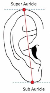
Figure 2. Auricle length is measured from the most superior to the most inferior projection. Both are based on using a horizontal line as the basis for the measurement.
This post was prefaced by stating that it would concentrate on two very basic measurements of the human auricle – those of auricle length and auricle width.
Many different methods are used to measure the surface features of the human auricle. Because of this, measurement comparisons are frequently difficult to make. But of all the measurements, that which is most likely to be measured the same way is that of auricle length (Figure 2).
Figure 2. Auricle length is measured from the most superior to the most inferior projection. Both are based on using a horizontal line as the basis for the measurement.
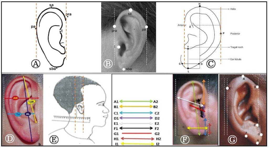
The width (breadth) auricle measurement shows among the greatest variability in procedure, as shown in Figure 3. It is because of differences in the width measurement reference points that result in an inability to directly compare measurements from certain studies.

Figure 3. Illustrations of methods used to measure the width of the auricle.
(A) Ferrario et.al. (1999) measurement of width. The width is measured as a horizontal line measured between the two vertical dotted lines that this author added. PRA is the area where the auricle helix meets the scalp superiorly. PA is the posterior-most length where it approximates a vertical line.
(B) Sforza et. al. (2009), following a suggested method of Farkus, appears to be the most accepted method, as a horizontal distance from pra to pa.
(C) Doepa et. al. (2013) appear to use the same method as (B), but for some reason have the horizontal line drawn shorter than were the auricle meets the scalp.
(D) Tatlisumak et. al. (2015) appear to use method (B) for the width measurement.
(E) Shireen et. al. (2015) use a somewhat different measurement, using the most anterior and the most posterior points of auricle. This may or may not coincide with method (B) because the anterior location is not identified. This could be a forward-facing lobule.
(F) Kapil et. al. (2014) provide a rather unusual illustration in that the width runs from the tragus to the what appears to be the most posterior portion of the auricle. This is not based on a horizontal line measurement, but a point-to-point measurement. The author has proved the white dashed lines to represent what are considered to be the anterior and posterior landmarks ordinarily utilized.
(G) Eboh (2013) appears to measure the auricle width from the auricle/scalp landmark, but then measure horizontally across to where the line meets the helix, not a vertical line representing the most posterior part of the auricle.
Auricle Height and Width Measurements
Figures 4 and 5 provides graphs of auricle height and width measurements taken from various studies. An attempt was made to provide data for populations between approximately 17 and 30 years of age. A later post will provide information about how the auricle height and width measure as a function of age.

Figure 4. Auricle height in mm for the studies identified below each bar, for male (blue), female (orange), and for a few studies that provided only combined male and female results (green). In the reference below each bar the study is identified, along with the n of the population, and age range.
The auricle height plotted for males is from 59.5 to 68.5 mm, a range of 9 mm. That for females has a smaller range (6.2 mm). There are not an equal number of studies because some studies were of males only. The studies that only provided combined data of males and females had a range of 5.2 mm. Interestingly, for those who use KEMAR (Knowles Electronics Manikin for Acoustical Research), it is noticed that what we identify as the small KEMAR ear (developed from data of Burkhard and Sachs in the graphs), was the longest of the studies for both males and females (one female studied tied it). This makes one wonder about the accuracy of the anatomical representation of the large KEMAR ear.

Figure 5. Auricle width in mm for the studies identified below each bar, for male (blue), female (orange), and for a few studies that only combined male and female results (green). In the reference below each bar the study is identified, along with the n of the population, and age range.
Auricle width comparisons are shown in Figure 5, with male ears showing a 12 mm range. Female auricle widths show a slightly smaller range of 9.8 mm. The results of the combined gender data show an estimated average width closer to the female, rather than the male widths. The width identified with the KEMAR small ear (Burkhard and Sachs in the graph) still measures to be closer to the greatest widths measured.
Summary
Auricle height and width measurements show considerable variability both between genders, and also within genders. This will be addressed in a later post.
References
- Sullivan, RF. Forum: Sullivan and Sullivan, Inc., Garden City, NY. https://www.rcsullivan.com/www/ears.htm – 13.0, 1996.Abeysekera JDA, Shahnavaz H. (1989). Body size variability between people in developed and developing countries and its impact on the use of imported goods. Int J Ind Ergon 4:139-149.
- Brucker MJ, Patel J, Sullivan PK (2003). A morphometric study of the external ear: age and sex-related differences. Plast Reconstr Surg 112 (2):647-652.
- Gualdi-Russo E. (1998). Longitudinal study of anthropometric changes with aging in an urban Italian population. Homo 49: 241-259.
- Ferrario VF, Sforza C, Ciusa V, Serrao G, Tartaglia GM. (1999). Morphometry of the normal ear: a cross-sectional study from adolescence to mid-adulthood. J Craniofac Genet Dev Biol 19 (4):226-233.
- Sforza C, Grandi G, Binelli M, Tommasi DG, Rosati R, Ferrario VF. (2009). Age- and sex-related changes in the normal human ear. Forensic Sci Int 187 (1): 110.e1-7.
- Purkait R, Singh P. (2007). Anthropometry of the normal human auricle: a study of adult Indian men. Aesthetic Plast Surg 31 (4):371-379.
- Deopa D, Thakkar HK, Prakash C, Niranjan R, Barua MP. (2013). Anthropometric measurements of external ear of medical students in Uttrarakhand Region. J Anat Soc India. 62: 79-83.
- Saha, PN. (1985). Anthropometric characteristics among industrial workers in India. Proceedings of International Symposium on Ergonomics in Developing Countries, Jakarta, Indonesia. 158-161.
- Kalcioglu MT, Miman MC, Toplu Y, Yakinci C, Ozturan O. (2003). Anthropometric growth study of normal human auricle. Int J Pediatr Otorhinolaryngo 67 (11): 1169-1177.
- Kalcioglu MT, Toplu Y, Ozturan O, Yakinci C. (2006). Anthropometric growth study of auricle of healthy preterm and term newborns. Int J Pediatr Otorhinolaryngol 70 (1): 121-127.
- Barut C, Aktunc E. (2006). Anthropometric measurements of the external ear in a group of Turkish primary school students. Aesthet Plast Surg 30 (2): 244-259e.
- Wang B, Dong Y, Zhao Y, Shizhu BS, Wu G. (2011). Computed tomography measurement of the auricle in Han population of north China. J Plast Reconstr Aesthetic Surgery 64 (1):34-40.
- Azaria R, Adler N, Silfen R, Regev D, Hauben DJ. (2003). Morphometry in the adult human earlobe: a study of 547 subjects and clinical application. Plast Reconstr Surg 111 (7): 2398-23402.
- Jung SH, Jung SH. (2003). Surveying the dimensions and characteristics of Korean ears for the ergonomic design of ear-related products. Int J Ind Ergon (31):361-373.
- Ekanem AU, Garba SH, Musa TS, Dare ND. (2010). Anthropometric study of the pinna (auricle) among adult Nigerians resident in Maiduguri metropolis. J Med Sci 10 (6): 176-180.
Graph References
- Burkhard MD, Sachs RM. (1975). Anthropometric manikin for acoustic research. Journal of the Acoustical Society of America, (58): 214-222.
- Dreyfus, H. (1967). The Measure of Man (Whitney Library of Design, New York).
- Tatlisumak E, Yavus MS, Kutlu N, Asirdizer M, Yoleri L, Aslan A. (2015). A symmetry, handedness and auricle morphometry. J. Morphol., 33(4): 1524-1548.
- Singh P, Purkit R. (2006). Anthropological study of human auricle. JIAFM, 28 (2):46-48.
- Ferrario VF, Sforza C, Ciusa V, Serrao G, Tartaglia GM. (1999). Morphometry of the normal ear: a cross-sectional study from adolescence to mid-adulthood. Craniofac. Genet. Dev. Biol. 19 (4):226-233.
- Shireen S, Karadkhelkar VP. Anthropometric measurements of human external ear. (2015). Journal of Evolution of Medical and Dental Sciences, Vol. 4, Issue 59, July 23; Page: 10333-10338, DOI: 10.14260/jemds/2015/1489.
- Sforza C, Grandi G, Binelli M, Tommasi DG, Rosati R, Ferrario VF. (2009). Age- and sex-related changes in the normal human ear. Forensic Sci. Int. 187 (1): 110.e1-7.
- Deopa D, Thakkar HK, Prakash C, Niranjan R, Barua MP. (2013). Anthropometric measurements of external ear of medical students in Uttrarakhand Region. J Anat Soc India. 62: 79-83.
- Bozkir MG, Karakas P, Yavus M, Dere F. (2006). Morphometry of the external ear in our adult population. Aesthetic Plast. Surg., 30(1):81-85.
- Kapil V, Bhawana J, Vikas K. (2014). Morphological variation of ear for individual identification in forensic cases: a study of an Indian population. Research Journal of Forensic Sciences, 2(1): 1-8, January.
- Algazi, V.R, Duda, R.O., Thompson, D.M., Avendano, C. (2001). The CIPIC HRTF Database, IEEE Workshop on Applications of Signal Processing to Audio and Acoustics, 2001.
Brucker MJ, Patek J, Sullivan PK. (2003). A morphometric study of the external ear: age- and sex-related differences. Plast Reconstr Surg. 112(2): 647-652.


