
Just imagine, you wake up one morning and the entire room is spinning around you. You have no idea why this is happening or how to make it stop. You notice that if you remain still the spinning will stop, but whenever you move again, the spinning sensation starts again. Because the world appears to be spinning around, you become nauseous, sweaty, and perhaps even vomit. Now let’s imagine that this scenario could recur when you move for up to months in duration. This would be a terrifying scenario for anyone experiencing it, and for many these symptoms can lead to a sense of hopelessness.
The scenario described to you in the previous paragraph would be enough to frighten even the most unshakable individual and is more common than one might think. The false sensation of the world spinning or moving, even while remaining still, is a sensation known as vertigo.
Are Vertigo and Dizziness the Same thing?
The term vertigo is one that most individuals have heard of, but all too often is misused to describe any dizziness symptom, often times leading to an incorrect diagnosis or no diagnosis at all. A sensation of vertigo falls under the more general term of dizziness.
The term dizziness can be used to describe many sensations including: vertigo, lightheadedness, imbalance, disorientation, blurred vision, as well as intolerance to visual or physical motion.
A more specific description of dizziness can dictate what assessment and treatments are implemented. In order to better understand what vertigo is, as well as ultimately what is causing it and how to treat it, using the correct terminology is essential.
What Causes Vertigo?
Vertigo has a multitude of potential causes, but the majority of the time it is due to just a handful of the most common disorders. The number one reason for experiencing vertigo is due to a vestibular disorder.
Vestibular disorders are typically non-life-threatening and largely consist of peripheral vestibular (ear) disorders; however, there are central vestibular (brain) disorders that exist that can be more worrisome.
Peripheral vestibular disorders are some of the most common causes of dizziness, with studies showing 32-54% of all dizziness in a clinical setting being attributed to peripheral vestibular (ear) disorders. 3,8,11 It has been shown that up to 90% of patients with a symptom of vertigo potentially have vestibular disorders.3
There are also non-vestibular causes of vertigo, but the primary focus of this article will be related to vestibular disorders as they are relatively common.
Peripheral Vestibular Disorders
For those not familiar, the peripheral vestibular system includes the inner ear and consists of both angular (head turn) and linear acceleration (forward/backward & up/down) sensors. These sensors allow for visual stability during head movement by sending reflexive signals to the eyes, causing the eyes move in the opposite direction of the head. These sensors also contribute to reflexive postural control by sending information to the lower extremities for maintenance of balance.
The angular sensors are called semicircular canals and the linear sensors are the otolith organs. Disorders of the peripheral vestibular system are not directly life-threatening but can produce intense vertiginous symptoms, disequilibrium, disorientation, and are often times accompanied by nausea and vomiting.
Central Vestibular Disorders
Central vestibular disorders refer to an abnormality of the brain or the nerve pathways between the inner ear and the brain. In general, structural central vestibular disorders such as strokes or tumors are far less common, but more worrisome than peripheral vestibular disorders.
Vestibular migraine or migraine associated vertigo, while technically considered a central vestibular disorder, is the exception, as it is very common but is not life threatening.
Peripheral Vestibular Disorders:
Benign Paroxysmal Positional Vertigo (BPPV)
The most common peripheral vestibular disorder is the condition of Benign Paroxysmal Positional Vertigo (BPPV), accounting for anywhere from 17-42% of all patients seen with symptoms of vertigo.2
The condition of BPPV causes brief episodes of vertigo provoked by a head movement or position change. With the condition of BPPV, the episode of vertigo typically resolves within one minute following the provoking head movement. In between the brief episodes of vertigo, one can be left with a sense of disequilibrium or disorientation. Common head movements that may make someone with BPPV experience intense vertigo are rolling over in bed, tilting one’s head backward or forward, or upon first lying down in bed.
BPPV is caused when otoconia (calcium carbonate crystals), that are supposed to be on top of the linear vestibular sensors (otoliths), migrate into one of the semi-circular canals. When the individual moves their head, the otoconia move within the canal that they have migrated into, and this asymmetrically stimulates the inner ears creating an involuntary eye movement called nystagmus. The sensation of vertigo is due to the nystagmus. The nystagmus will persist until the otoconia come back to rest following the provoking head movement, which typically is less than one minute. While bothersome, this condition is benign, and for many, will resolve over a period of several months spontaneously.2
Assessment of BPPV
Due to how common BPPV is, a number of healthcare professions assess and treat for it, including but not limited to: physicians, audiologists, physical therapists, and occupational therapists.
BPPV is assessed clinically through the introduction of position change so that any displaced otoconia will be set into motion and will provoke an episode of positional vertigo. Common provoking movements include laying the individual on their back with their neck extended backward and/or turning the individual’s head side to side while they are lying down.
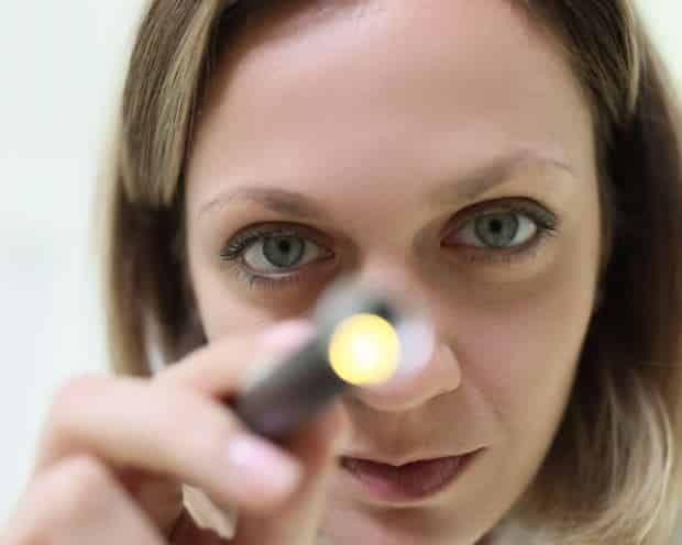 A trained clinician can tell by the pattern of nystagmus which semicircular canal has the otoconia within it, and prescribe the correct repositioning maneuver. It is important to note that even if the clinician does not trigger an episode of vertigo with position change during examination, this does not rule out the condition of BPPV. The condition of BPPV does not cause an episode of vertigo every time the individual changes position and often times cannot be reliability triggered in a clinical setting.
Treatment of BPPV
There are effective treatments for this condition called canalith repositioning maneuvers (more commonly known as the Epley or Semont maneuvers) that have been shown to greatly expedite the recovery process.
These canalith repositioning maneuvers consist of a series of timed head movements in certain positions designed to remove the offending otoconia from the semicircular canal. Canalith repositioning has been shown to be up to 98% effective when performed correctly for posterior semicircular canal BPPV, which accounts for 85-95% of all BPPV.2 Other repositioning maneuvers may be required if other semicircular canals are affected.
Following successful canalith repositioning or spontaneous resolution of the symptoms of BPPV, most individuals are asymptomatic; however, it is important to note that the chance of recurrence of BPPV has been shown to be as high as 36%.2 Also, if someone has another underlying peripheral vestibular disorder that has caused damage to the ear, they will likely not have complete symptom resolution. This demonstrates the need for further clinical evaluation if ones symptoms of BPPV are not resolved following canalith repositioning.
In fact, we often tell patients “these symptoms should get better with time, not worse”, to demonstrate the need for further assessment if one’s symptoms do not resolve with canalith repositioning and/or time.
Vestibular Neuritis/Labyrinthitis
The second most common peripheral vestibular disorder is what is known as a vestibular neuritis or labyrinthitis. This condition has been shown to affect around 7% of those seen in a clinical setting.1
Vestibular neuritis is thought to be the result of a viral inflammation of the vestibular nerve that either temporarily or permanently disrupts nerve transmission between the inner ear and the brain. This reduction in ear input to the brain creates an asymmetry in vestibular input and results in nystagmus (jerking eye movement), creating a sensation of vertigo. The nystagmus created from a vestibular neuritis is more persistent than what is seen in BPPV, and will typically be present for hours to days, resulting in a sensation of vertigo for this same time period.
The constant and intense sensation of vertigo for that extended period of time can lead to co-occurring disequilibrium, as well as significant nausea and vomiting.
The sensation of vertigo due to a vestibular neuritis will be present even if the affected individual is remaining completely still because it is the reduced vestibular input to the brain causing the sensation. Over time, the individual’s brain can make sense of the vestibular asymmetry through a process called “compensation”, which is basically a brain recalibration process, reducing the symptoms associated with the vestibular asymmetry. In other words, the brain gets used to the difference between the two ears, and the vertigo decreases.
The key distinction between vestibular neuritis and labyrinthitis is that a vestibular neuritis typically only affects the vestibular portion of the inner ear, while a labyrinthitis also affects the hearing portion of the inner ear, the cochlea.15 Therefore, those with a labyrinthitis could have decreased hearing, tinnitus (noises) or a sensation of pressure in the affected ear. With a vestibular neuritis, some individuals have complete recovery of function and are thus asymptomatic after a period of time, while others can be left with varying degrees of damage in the affected ear. For those with significant damage or with no function at all left in the remaining ear, there can be more persistent, often lifelong symptoms.
Some of the more common symptoms could include disorientation with quick head movements towards the affected ear and imbalance in scenarios with poor lighting or when walking on uneven ground.

Assessment of Vestibular Neuritis/Labyrinthitis
An audiologist, an individual with either master’s or doctoral level clinical training in diagnostics of the ear, typically assesses for this condition.
Vestibular neuritis and labyrinthitis are frequently assessed with a test battery called a VNG (videonystagmography) and a hearing evaluation. Other tests to fully understand the degree of vestibular dysfunction and the degree of compensation/recalibration include: rotational chair, video head impulse test (vHIT), and vestibular evoked myogenic potential (VEMP) to name a few.
These tests are not, however, routinely performed in many clinics due to the costs associated with the equipment and the level of training of the staff. It is essential to understand that an abnormal VNG showing peripheral vestibular dysfunction can be used to make a diagnosis of a weakness in one or both ears, but that a VNG can miss some peripheral vestibular disorders, as it only assesses one aspect of vestibular function.
Treatment of Vestibular Neuritis/Labyrinthitis
Typically, medical treatment such as prescribing medications for this condition is performed by a physician or specialty provider such as an Otolaryngologist, a physician with specialty in disorders of the ears, nose and throat. In the initial acute stages of a vestibular neuritis, treatment often entails the use of medications to help reduce nausea and/or anxiety associated with symptoms.
Many individuals who have suffered permanent vestibular damage from a vestibular neuritis or labyrinthitis can compensate for this injury with regular daily physical activity, but some may require specialized vestibular rehabilitation designed to promote the compensation process from vestibular injury. Those who require more specialized therapy will often see a physical therapist with training in the rehabilitation of vestibular injuries.
Meniere’s Disease
 Many individuals are familiar with another peripheral vestibular disorder called Meniere’s disease; however, the disease is not all that common. Recent studies have shown that Meniere’s disease affects less than 1% of patients with dizziness.1
There is some disagreement on the exact cause of Meniere’s disease, but for the most part, it is agreed upon that it is related to poor regulation of an inner ear fluid called endolymph. Traditionally it was thought that this could cause fluctuations in inner ear fluid levels in just the affected ear, which asymmetrically stimulated the ears, sending different signals to the brain from each inner ear. As discussed in other sections, when the ears send conflicting signals to the brain, nystagmus (involuntary jerking eye movement) is generated, thus creating a sensation of vertigo. Also, one typically has decreased hearing, ear pressure, and tinnitus (noises in the ear) in the affected ear with the episode of vertigo.
These episodes tend to last anywhere from minutes to hours in duration. Over a period of hours, the fluid levels within the inner ear were thought to stabilize, allowing for the vertigo to dissipate, the hearing to return to some degree, and the ear pressure and tinnitus to lessen. In recent years, this traditional view has been opposed by many other potential theories for the cause of Meniere’s disease. With repeated attacks of Meniere’s disease, the inner ear is left damaged, often resulting in a more chronic sense of disequilibrium, hearing loss, ear pressure, and tinnitus in the affected ear.
Assessment of Meniere’s Disease
An audiologist typically completes the assessment and an Otolaryngologist handles the treatment of Meniere’s disease. There is no single test for Meniere’s disease, so the diagnosis is typically made based on the patient’s symptoms and a pattern of diagnostic test findings that are not typical of other ear disorders.
The expert consensus committee recommends two categories for this diagnosis: definite and probable Meniere’s disease. For a diagnosis of definite Meniere’s disease, one should have episodes of vertigo for 20 minutes up to 12 hours, with documented low-frequency (low-pitch) sensorineural (inner ear) hearing loss in the affected ear. Also, there should be fluctuating hearing, tinnitus, and/or a pressure sensation in the affected ear.10 The category of probable Meniere’s disease requires the individual to have episodic dizziness symptoms associated with fluctuating hearing, tinnitus, and/or pressure in the affected ear, lasting anywhere from 20 minutes up to 24 hours.10

Meniere’s disease patients tend to show distinctive, low-frequency hearing loss patterns on their hearing exam results
Meniere’s disease tends to appear on a hearing test with a rather unusual, low-frequency inner ear hearing loss on the affected side, making a hearing test essential for the diagnosis of definite Meniere’s disease. Depending on the duration of this disease process, the ear may or may not be damaged following the attack. Due to this, the hearing in the affected ear can return to normal following the episodes in the early stages of the disease. To be more certain of the diagnosis, it is helpful to complete multiple hearing tests when symptomatic, in order to show potential fluctuations in hearing in the affected ear.
Similar to assessment for vestibular neuritis/labyrinthitis, other tests to fully understand the degree of vestibular dysfunction, as well as the individual’s degree of compensation (calibration) include: a test battery called a VNG (videonystagmography) rotational chair, video head impulse test (vHIT), and vestibular evoked myogenic potential (VEMP). Another frequently implemented test for Meniere’s disease is electrocochleography (ECOG), used to determine if there is abnormal fluid pressure within the ear.
Treatment of Meniere’s Disease
Treatment of Meniere’s disease most often begins with more benign, but often times less effective treatments. If these are ineffective at symptom control, then treatments that are more effective but with greater risk of adverse side effects are explored. Typical first line treatments include reducing ones intake of salt, caffeine, alcohol, and tobacco. A diuretic (fluid pill) may also be utilized.
If these treatments are not effective in symptom control, the clinician will typically move to more effective treatments, with these treatments often being more invasive and/or having a greater risk for adverse side effects. Some of these treatments include steroid injections into the middle ear or the injection of powerful antibiotics into the middle ear to deaden the inner ear vestibular nerve endings.
There are also surgical procedures such as building a drain to allow for fluid runoff from the inner ear, or cutting the vestibular nerve.
Central Vestibular Disorders
Vestibular Migraine
Vestibular migraine is the second most common cause of recurrent dizziness symptoms
Vestibular migraine is thought to be the most common form of central vestibular dysfunction, as well as the second most common cause of recurrent dizziness symptoms.1 Migraines in general are thought to be a vascular abnormality with problems in either the constriction and/or dilation of the blood vessels in the brain, resulting in a vasospasm.16
In vestibular migraine, this vasospasm occurs in the brain along the central vestibular pathways, disrupting the brains interpretation and integration of peripheral vestibular input, leading to a sensation of vertigo. An episode of vertigo due to vestibular migraine can last anywhere from minutes to days in duration. Dizziness related to vestibular migraine is typically spontaneous in nature but can also be provoked by certain foods, hormonal fluctuations or changes in weather. Recent studies show that vestibular migraine affects 6-9% of patients seen in specialized clinics. 1
Additionally, it has been reported that up to 38% of individuals with migraine headache also have episodes of vertigo, making vestibular migraine fairly prevalent. 12
When someone is having an episode of vertigo due to vestibular migraine, they will frequently have other migraine symptoms that occur in close proximity to the episode, including:
- Headache
- Visual aura
- Sensitivity to light and/or sound
A migraine visual aura is a visual disturbance typically consisting of partial vision loss, bright lights, or zig-zag lines in one’s vision.
Assessment of Vestibular Migraine
The current recommended criteria to reach a diagnosis of vestibular migraine suggests that the individual should have had multiple episodes, with dizziness symptoms that last for minutes to hours in duration, with associated migraine symptoms during at least 50% of the episodes. Also, there should be no other diagnosis that could explain the symptoms.9
Based on these criteria, it is essential to determine what symptoms go along with the episodes of vertigo. There is not a diagnostic test for migraine and in order for a diagnosis to be made, other potential causes must be ruled out. In this way, assessment for vestibular migraine is similar to the assessment performed for peripheral vestibular disorders, with the expectation that the majority of the vestibular testing should be within normal limits.
Assessments to rule out other causes for vertigo are typically completed by audiologists, otolaryngologists, and other physician providers.
Treatment of Vestibular Migraine
A Neurologist typically treats and diagnoses vestibular migraine. The typical first line treatments for vestibular migraine include lifestyle modification such as trying to determine and avoid potential dietary triggers and reducing stress.
If these lifestyle modifications are unsuccessful in controlling the symptoms, then medications are typically prescribed to reduce the frequency or severity of the episodes.
Vertebrobasilar (Brainstem) Stroke
Many individuals suffering from an episode of intense spontaneous vertigo minds immediately fear the worst-case scenario and think they could be having a stroke. While strokes do occur frequently, they are actually not one of the more common reasons someone has an episode of isolated vertigo.
Recent evidence shows that around 3% of patients presenting to the emergency department with general dizziness were due to a stroke and around 1% of those with specific symptoms of vertigo were due to a stroke.5
Assessment of Vertebrobasilar Stroke
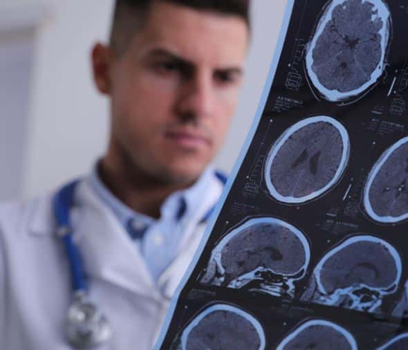 Most individuals assessed for stroke are seen by physicians in the emergency department. Examination for someone having a stroke often consists of a basic bedside neurological examination, and if the clinical suspicion is high, then computed tomography (CT) or magnetic resonance imaging (MRI) of the brain would likely be completed.
Traditionally, to reach a diagnosis of what was causing vertigo, worrisome, potentially life-threatening pathologies such as stroke were ruled out first. However, due to the majority of vertigo being attributed to benign peripheral vestibular disorders, this approach is relatively inefficient in reaching a timely diagnosis.7
In recent years, assessment techniques to quickly distinguish stroke from benign peripheral disorders have been brought forth.14 Also, these techniques combined with the implementation of positional testing to detect BPPV would likely improve efficiency in reaching a diagnosis for what is causing one’s vertigo.7
Non-Vestibular Vertigo:
Cardiovascular Disease
Cardiovascular disease encompasses a multitude of diagnoses and consists of any disorder involving the heart or blood vessels. Traditionally, cardiovascular disorders were thought to only cause symptoms of one feeling lightheaded or near faint. This assumption, however, is likely flawed as recent studies show that vertigo was present in 63% of patients with a primary cardiovascular diagnosis. The same study showed that for 37% of patients, vertigo was their only dizziness symptom.13

Orthostatic hypotension is thought to affect 10-15% of individuals seen clinically for all dizziness
A common type of cardiovascular related dizziness is a condition known as orthostatic hypotension. This condition occurs when one’s blood pressure drops when rising from a seated to standing position and causes brief symptoms of dizziness upon rising. The condition of orthostatic hypotension can be caused by medication, dehydration, anemia, low blood sugar, and much more. Orthostatic hypotension is thought to affect 10-15% of individuals seen clinically for all dizziness.11
There are a number of cardiovascular diagnoses that can cause dizziness and as shown above, just because an individual is experiencing a symptom of vertigo, it does not mean that a cardiovascular diagnosis is ruled out by any means.
These conditions are typically assessed and treated by a primary care physician and/or a cardiologist.
Psychiatric Conditions
A psychiatric condition refers to any condition that alters one’s emotions, thoughts, or behaviors and as such, encompasses a number of diagnoses. Psychiatric or psychogenic causes of dizziness are thought to be the primary cause in 2-6% of all dizziness and around 1% of cases of vertigo.3,8,11
It is important to note that purely psychiatric conditions, while not a common cause of vertigo, do often accompany vestibular disorders in the form of anxiety and depression due to the bothersome nature of vestibular disorders.1 These conditions are assessed and managed by a psychologist or psychiatrist.
When to Seek Help for Vertigo
Vertigo is relatively common, and most symptoms of vertigo are related to benign vestibular causes, but the only way to reach a diagnosis is to seek help. Anyone having symptoms of dizziness or vertigo should consult either their primary care provider or report to the emergency department if their symptoms are severe enough.
For those interested in learning more on comprehensive vestibular assessment click here.
References:
- Agrawal, Y., Ward, B.K., & Minor, L.B. (2013) Vestibular dysfunction: Prevalence, impact and need for targeted treatment. Journal of Vestibular Research. 23(3): 113-117.
- Battacharyya, N., Gubbels, S.P., Schwartz, S.R., Edlow, J.A., El-Kashlan, H., Fife, T. ……& Corrigan, M.D. (2017) Clinical practice guideline: Benign paroxysmal positional vertigo (update). Otolaryngology-Head and Neck Surgery. 156(3S): S1-S47.
- Bösner, S., Schwarm, S., Grevenrath, P., Schmidt, L., Hörner, K., Beidatsch, D. …..& Haasenritter, J. (2018) Prevalence, aetiologies and prognosis of the symptom dizziness in primary care – a systematic review. BMC Family Practice. 19(33): 1-13. DOI 10.1186/s12875-017-0695-0
- Drachman, D.A., & Hart, C.W. (1972) An approach to the dizzy patient. Neurology. 22(4): 323-334.
- Kerber, K.A., Brown, D.L., Lisabeth, L.D., Smith, M.A., & Morgenstern, L.B. (2006) Stroke among patients with dizziness, vertigo, and imbalance in the emergency department: A population-based study. Stroke. 37(10). 2484-2487
- Kerber, K.A., Callaghan, B.C., Telian, S.A., & Meurer, W.J. (2017) Dizziness symptom type prevalence and overlap: a US nationally representative study. The American Journal of Medicine. 130(12): 1465e1-1465e9.
- Kerber, K.A. & Newman-Toker, D.E. (2015) Misdiagnosing dizzy patients: common pitfalls in clinical practice. Neurologic Clinics. 33(3): 565-575.
- Kroenke, K., Lucas, C., Rosenberg, M., Scherokman, B., Herbers, J., Wehrle, P. &Boggi, J. (1992) Causes of persistent dizziness: A prospective study of 100 patients in ambulatory care. Annals of Internal Medicine. 117(11): 898-904. DOI: 10.7326/0003-4819-117-11-898
- Lempert, T., Olesen, J., Furman, J., Waterston, J., Seemungal, B., Carey, J…..& Newman-Toker, D. (2012) Vestibular migraine: diagnostic criteria. Journal of Vestibular Research. 22: 167-172.
- Lopez-Escamez, J.A., Carey, J., Chung, W.H., Goebel, J.A., Magnusson, M., Mandala, M. …. & Bisdorff, A. (2016) Diagnostic criteria for Menière’s disease. Consensus document of the Bárány Society, the Japan Society for Equilibrium Research, the European Academy of Otology and Neurotology (EAONO), the American Academy of Otolaryngology-Head and Neck Surgery (AAO-HNS) and the Korean Balance Society. Acta Otorrinolaryngol. 67(1): 1-7. doi: 10.1016/j.otorri.2015.05.005.
- Molnar, A., & McGee, S. (2014) Diagnosing and treating dizziness. Medical Clinics North America. 98: 583-596.
- Neuhauser, H.K., Leopold, M., Von Brevern, M., Arnold, G. & Lempert, T. (2001) The interrelations of migraine, vertigo, and migrainous vertigo. Neurology. 56:436–441.
- Newman-Toker, D., Dy, F., Stantond, V., Zee, D., Calkins, H. & Robinson, K. (2008). How often is dizziness from primary cardiovascular disease true vertigo? a systematic review. J Gen Intern Med 23(12): 2087–2094.
- Newman-Toker, D., Kattah, J., Talkad, A., Wang, D., & Hsieh, Y. (2009) HINTS to diagnose stroke in the acut vestibular syndrome-three step bedside oculomotor exam more sensitive than early MRI DWI. Stroke. 40(11) : 3504-3510.
- Silvoniemi P. (1988) Vestibular neuronitis: an otoneurological evaluation. Acta Otolaryngology. 453:1-72.
- Thompson, T.L. & Amedee, R. (2009) Vertigo: a review of common peripheral and central vestibular disorders. The Ochsner Journal. 9: 20-26

Brady Workman, AuD, is an audiologist in the Balance Disorders program at Wake Forest Baptist Health Center. He has authored several articles relating to balance and vestibular disorders as a regular contributor and co-editor of the Dizziness Depot at Hearing Health & Technology Matters. Brady received his doctorate of audiology from East Tennessee State University in 2018 and is licensed by the North Carolina Board of Examiners for Speech Language Pathologists and Audiologists and is a fellow of the American Academy of Audiology.