
AO Technologies, a startup founded by researchers at the University of Southern California (USC), has received an award of up to $3.7 million from the Advanced Research Projects Agency for Health (ARPA-H) to further develop a device designed to image the living inner ear.
The funding will support continued development of the company’s cochleoscope, an imaging platform intended to provide otolaryngologists with detailed views of the cochlea that could improve the diagnosis and management of inner ear disorders.
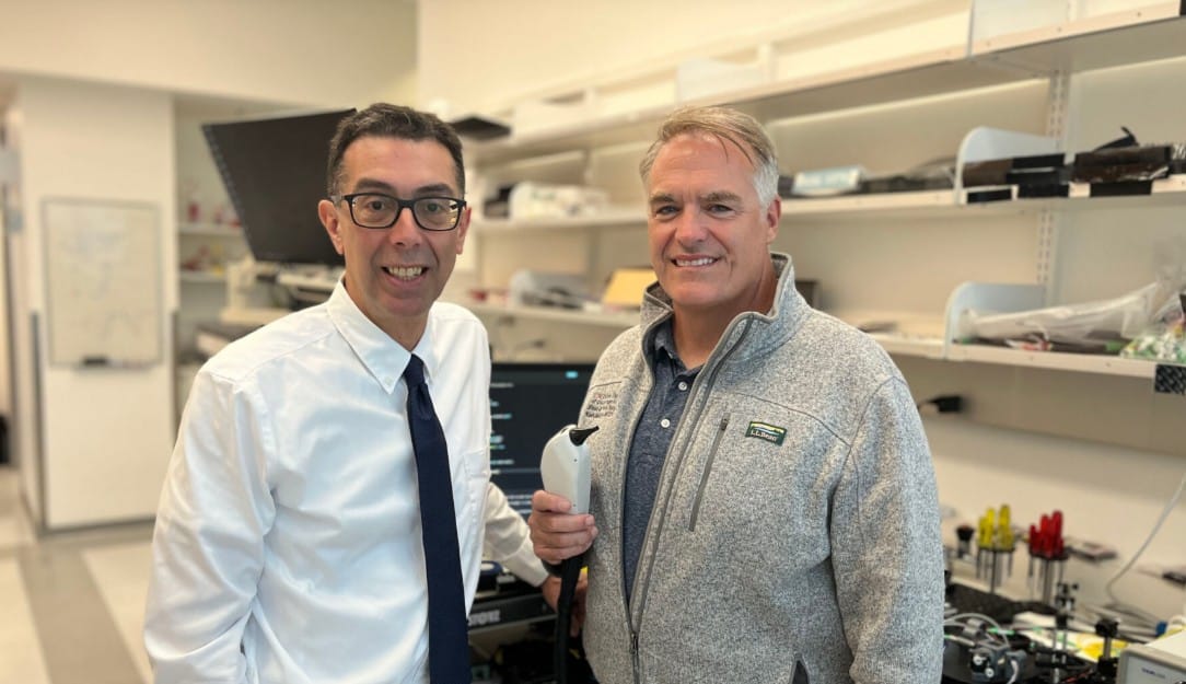
The technology is being developed by John Oghalai, MD, Chair of the USC Caruso Department of Otolaryngology – Head and Neck Surgery, and Brian Applegate, PhD, Professor of Otolaryngology – Head and Neck Surgery, Biomedical Engineering, and Electrical and Computer Engineering at USC.
“Our goal is to have something that we can put into the ear that can image the tissues of the cochlea, which, in humans, is encased in the densest bone in the body,” said Oghalai.
Addressing a Longstanding Inner Ear Imaging Challenge
Unlike many other parts of the body, the cochlea is enclosed within dense temporal bone, making it extremely difficult to visualize without invasive procedures. While optical coherence tomography (OCT) has shown promise in research settings, producing clinically useful images through bone has remained a significant technical challenge.
According to the researchers, the cochleoscope combines advances in optical hardware with new imaging algorithms to improve image quality while maintaining a form factor suitable for clinical use.
“Our first set-up was as big or bigger than a Toyota Corolla and weighed about as much,” Oghalai said. “Now the equipment fits on a medical cart in a clinical setting.”
Oghalai and Applegate have collaborated on inner ear imaging research for nearly two decades, beginning with animal studies that explored the use of OCT to visualize cochlear structures. Their work eventually led to the formation of AO Technologies to help translate the technology into a clinical device.
The latest funding builds on years of research aimed at making OCT practical for imaging the living inner ear. HHTM has previously reported on Oghalai’s NIH-supported work to develop OCT-based cochlear imaging, as well as USC research using OCT to visualize fluid movement within the inner ear.
ARPA-H Award Supports Clinical Translation
The new award will fund continued refinement of the cochleoscope over the next four years, with the goal of evaluating the technology in patients and improving its clinical utility.
The project builds on previous support from the National Institutes of Health, USC’s Stevens Center for Innovation, and the Nemirovsky Engineering and Medical Opportunity (NEMO) Prize, which helped advance early-stage development of the technology.
“The NEMO Prize enabled us to try some experiments that would have been considered very risky by a traditional funding agency,” Applegate explained.
Rather than relying on venture capital financing, AO Technologies has largely advanced through federal grants and university-supported commercialization programs.
“The startup very much spun out from the university, and they have been helping us along the way to make sure we’re successful,” Applegate said. “Venture capital wants you to make a profit tomorrow, but we have the ability to boot-strap our company without those external pressures.”
Because the startup is closely connected to USC, the researchers say they are able to continue working with university labs that are already equipped to refine and test the device.
“Of course, we have to manage conflicts of interest according to USC policy,” said Oghalai, “but the labs at the university that are set up for this research are the best way we know to iteratively improve the device and test new technology.”
Imaging Emerging as a Key Need for Inner Ear Therapeutics
The award comes as interest in inner ear therapeutics continues to accelerate. Pharmaceutical and biotechnology companies are advancing investigational therapies for hearing loss, tinnitus, Ménière’s disease, and other inner ear disorders, yet clinicians currently have limited ability to directly visualize the microscopic structures those therapies are intended to treat.
Today, diagnosis of many inner ear disorders relies on a combination of audiometry, vestibular testing, patient symptoms, MRI, CT, and other clinical assessments. However, these tools do not directly visualize cochlear soft tissues at high resolution in living patients.
A clinically viable cochleoscope could eventually provide physicians with new insight into disease processes, help identify patients most likely to benefit from emerging therapies, and potentially allow clinicians to monitor treatment response over time.
Over the next four years, Oghalai and Applegate will work with ARPA-H to continue refining the cochleoscope, with the goal of producing images that can provide useful information for both patients and physicians.
“One of USC’s greatest strengths is that it is a collaborative institution,” Oghalai said. “The leadership facilitates that, and this is a great example.”
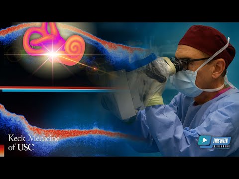
Editor’s note: HHTM has previously covered Dr. John Oghalai’s work on OCT imaging of the inner ear, including research focused on cochlear imaging and inner ear fluid dynamics. Readers can also watch HHTM’s 2025 interview with Dr. Oghalai on how OCT imaging could transform the diagnosis and management of Ménière’s disease and other inner ear disorders.
Source: USC
*featured image showing John Oghalai and Brian Applegate with the cochleoscope (Photo by Cristy Lytal) credit: USC